


Introduction
The mucosal immune system of the oropharyngeal cavity, which is the opening gate for digestive and respiratory systems, plays a crucial role in preventing pathogen entry into the human body [1]. The oral cavity is a suitable environment for microbial colonization; 392 taxa with approximately 700 species and 1500 microbial genomes have been identified as residing therein. Firmicutes, Actinobacteria, Proteobacteria, Fusobacteria, Bacteroidetes, and Spirochaetes constitute 96% of all oral bacterial phyla [2, 3]. Additionally, 85 fungal genera are found in the oral cavity [4]. Oral microbes colonize the oral cavity as a biofilm, which regulates oral homeostasis, oral immunity, food digestion, detoxification, inflammatory processes and is involved in disease prevention [5, 6].
A healthy oral environment consists of multiple symbiotic microbiota. Dewhirst et al. demonstrated major oral microbial phylum were Firmicutes (36.7%), Bacteroidetes (17.3%), Proteobacteria (17.1%), Actinobacteria (11.6%), Spirochaetes (7.9%), Fusobacteria (5.2%) [7]. However, poor oral hygiene may cause oral microbiota dysbiosis, which leads to dental bacterial plaque, gingivitis, and periodontitis [8]. In periodontitis, an outgrowth of pathogenic bacteria occurs, among which Actinobacillus actinomycetemcomitans, Porphyromonas gingivalis, and Fusobacterium nucleatum have been reported to be highly related to periodontal disease pathogenesis [9, 10]. The World Health Organization (WHO) estimated that approximately 20%–50% of the global population has periodontitis [11]. Patients with periodontitis are at high risk of developing stroke, peripheral artery disease, and coronary heart disease [12]. The WHO suggested that strategies for periodontitis prevention include practicing oral hygiene, having a healthy diet, using fluoride and antimicrobial agents, and smoking cessation [13]. Studies have shown that live probiotic strains inhibit oral pathogens and are the rationale for periodontal treatment; they are similar to antibiotics but without the major concern of antimicrobial resistance [14]. However, manufacturing viable probiotics is not feasible owing to challenges regarding preservation and viability stabilization [15]. Metabolites of viable probiotic strains (postbiotic) such as 10-Hydroxy-cis-12-octadecenoic acid and heat-inactivated probiotics have shown the potential to alleviate the disruption of the gingival epithelial barrier caused by periodontitis [16].
In 2019, the panel of International Scientific Association for Probiotics and Prebiotics (ISAPP) defined the term ‘postbiotics’ as a preparation of inanimate microorganisms and/or their components that confers a health benefit on the host [17]. Postbiotics are often considered as metabolites secreted by probiotic strains during fermentation and consist of microbial cell fractions, polypeptides, peptidoglycan-derived muropeptides, bacteriocins, peroxides, pili-type structures, short-chain fatty acids, teichoic acid, folate, vitamins, lactic acid, and extracellular polysaccharides. Probiotic components benefit human health, provide nutritional support, competitively inhibit pathogenic bacteria, and regulate the immune system [18, 19]. In addition, the fermentation products of lactic acid bacteria (postbiotics) have a unique flavor and beneficial nutrients and are therefore widely used in the food industry [20]. An in vitro study demonstrated that Lactobacilli postbiotics reduce colonization levels of A. actinomycetemcomitans, which are related to periodontitis [21]. However, clinical evidence proving that postbiotics reduce oral pathogenic bacteria and improve oral health is lacking. Additionally, our previous study revealed that certain heat-killed probiotics, including L. salivarius subsp. salicinius AP-32 and L. paracasei ET-66, effectively limit the growth of oral pathogenic bacteria in vitro [22]. Current clinical data indicate that oral lozenges made of viable strains, including L. salivarius subsp. salicinius AP-32, L. paracasei ET-66, and L. plantarum LPL28, can increase beneficial microbiota in the oral cavity, reduce the colonization of periodontitis-related bacteria, and increase the levels of salivary immunoglobulin A (IgA) [23].
Based on previous in-vitro screening of viable probiotic strains for improving oral health, we further investigated whether heat-killed probiotic (ET-66 and AP-32), and postbiotic lozenges (LPL28, ET-66, and AP-32) modulate oral microbiota, inhibit oral infectious pathogens, and change salivary IgA levels [22, 23]. The results can be applied in the production of supplementary foods for clinical oral health care in future.
Results
Postbiotics of AP-32, ET-66, and LPL28 strains showed effective bactericidal effects on oral pathogens S. mutans, P. gingivalis, F. nucleatum subsp. polymorphum, and A. actinomycetemcomitans
The experimental design was revealed in supplementary data (Supplementary Figure 1). First, we generated the fermentation products of AP-32, ET-66, and LPL28 as postbiotic oral lozenge, and examined its antipathogenic activity against oral pathogens (Figure 1). Compared with the postbiotic of a commercially available strain (LGG), the fermentation products (postbiotics) of AP-32, ET-66, and LPL28 strains had stronger bactericidal effects on oral pathogens, particularly S. mutans and A. actinomycetemcomitans (Figure 1A). The inhibition rates of S. mutans were significantly higher with the use of the postbiotics of AP-32 (62.6%), ET-66 (99.86%, p < 0.001), and LPL28 (94.35%, p < 0.001) than with the use of LGG postbiotic (41.64%). In addition, the inhibition rates of A. actinomycetemcomitans were significantly higher with the use of the postbiotics of AP-32 (17.92%, p < 0.05), ET-66 (77.86%, p < 0.001), and LPL28 (24.28%, p < 0.001) than with the use of LGG postbiotic (13.69%). All postbiotics effectively inhibited periodontal pathogens P. gingivalis BCRC 17689, P. gingivalis BCRC 17688, and F. nucleatum subsp. polymorphum (Figure 1A). LPL28 postbiotics had a higher inhibition rate of P. gingivalis BCRC 17688 (100%, p < 0.05*) than did LGG postbiotic (96.83%).
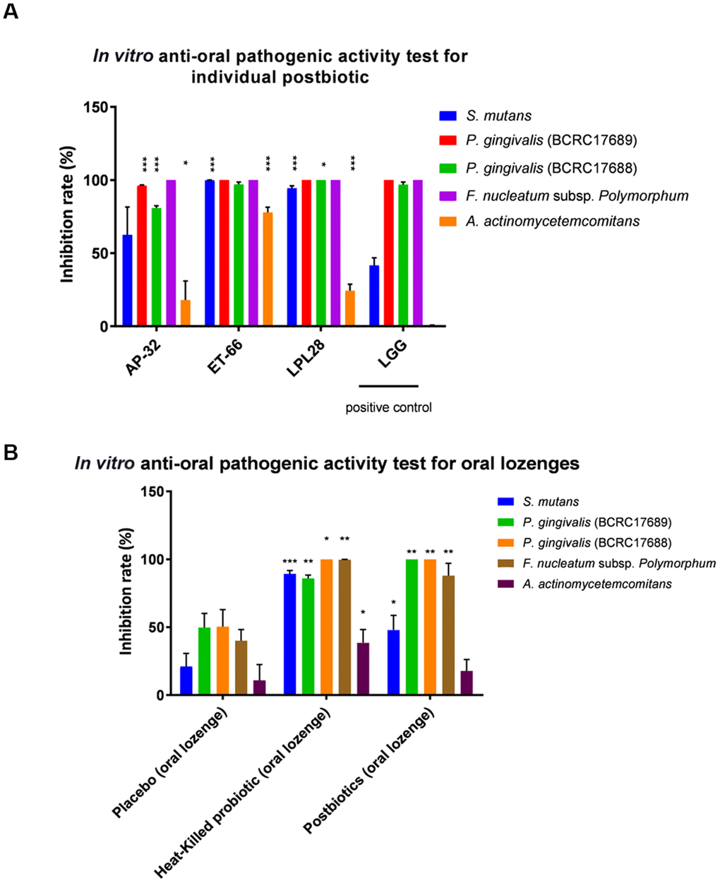
Figure 1. In vitro test for determining the antipathogenic activity of (A) individual postbiotic and (B) probiotic oral lozenges against oral pathogens. (A) Postbiotics of AP-32, ET-66, and LPL28 showed strong antibacterial activities compared with the positive control of LGG postbiotic. (B) Heat-killed AP-32 and ET-66 were used as inactivated probiotics, whereas metabolites of AP-32, ET-66, and LPL28 were used as postbiotics. *p < 0.05, **p < 0.01, and ***p < 0.001 compared with the positive control group (LGG postbiotic) or the placebo group (without the postbiotic). Data are presented as mean ± SD.
Postbiotic and heat-killed probiotic lozenges were effective, demonstrating in vitro bactericidal ability against oral pathogens
We prepared two oral lozenges, one with the postbiotics of AP-32, ET-66, and LPL28 and the other with heat-killed AP-32 and ET-66 probiotics for an in vitro bactericidal test before launching clinical trials (Figure 1B). The postbiotic oral lozenge group showed a significant increase in the inhibition rates of S. mutans, P. gingivalis (BCRC 17689), P. gingivalis (BCRC 17688), F. nucleatum, and A. actinomycetemcomitans, namely increases of 47.97% (p < 0.05, placebo = 21.02%), 100% (p < 0.01, placebo = 49.74%), 100% (p < 0.01, placebo = 50.38%), and 87.88% (p < 0.01, placebo = 39.96%), respectively, compared with the placebo group. The inhibition rate of A. actinomycetemcomitans in the experimental group slightly increased without a significant difference compared with the placebo group (oral lozenge: 17.78%; placebo: 10.85%). The heat-killed probiotic group significantly inhibited S. mutans, P. gingivalis (BCRC 17689), P. gingivalis (BCRC 17688), F. nucleatum, and A. actinomycetemcomitans by 89.32% (p < 0.001), 85.91% (p < 0.01), 95.32% (p < 0.05), 91.75% (p < 0.01), and 48.46% (p < 0.05), respectively.
Postbiotic and heat-killed probiotic lozenges effectively reduced pathogenic colonies in the saliva samples of participants
The 75 selected participants were randomly assigned to three groups: placebo, postbiotic lozenge, and heat-killed probiotic lozenge. We collected saliva samples at weeks 0, 2, and 4 after oral lozenge intake initiation and measured changes in their microbiota. Plaque weight was 0.37 ± 0.16 g at week 0, and the initial S. mutans in saliva (CFUs/mL) was 4.25E+06 ± 2.90E+06. The postbiotic lozenge significantly reduced the oral S. mutans bioburden to 60% (median) at week 4 (compared with the postbiotic lozenge at week 0 and placebo at week 4; p < 0.05 for both), and the heat-killed probiotic lozenge significantly reduced S. mutans to 61% (median) at week 4 (compared with the probiotic lozenge at week 4, p < 0.05; Figure 2A). Plate Count Agar (PCA) is a common microbiological growth medium used to monitor total viable bacterial populations of a sample [24]. The PCA agar plate was used to analyze the total bacterial population in the oral cavity. The result indicated that administrating postbiotic lozenges significantly decreased the total bacterial load to 98% at week 2 (compared with placebo at week 2, 160%, p < 0.05; Figure 2B) and to 104% at week 4 (compared with placebo at week 4 [187%] and postbiotic lozenge at week 0; p < 0.05 for both).
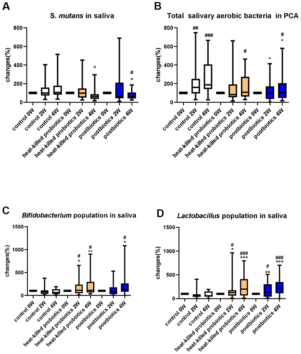
Figure 2. Microbial change (%) in saliva samples. Change (%) in the population of (A) S. mutans, (B) total bacteria, (C) Bifidobacterium, and (D) Lactobacillus in participants’ saliva at 0, 2, and 4 weeks of oral lozenge intake. The oral lozenges contained postbiotics or heat-killed cells. Participants in the control group consumed placebo lozenges without the postbiotic content (*p < 0.05, **p < 0.01, and ***p < 0.001 compared with the control group; #p < 0.05, ##p < 0.01, and ###p < 0.001 in reference to the values at week 0). Data are presented as medians (n = 25 in each group).
Postbiotic and heat-killed probiotic lozenges effectively increased beneficial microbial strains in saliva samples
The change in the Bifidobacterium population in the participants’ oral cavity was further measured after oral lozenge intake. The results revealed that the postbiotic oral lozenge significantly increased the Bifidobacterium population to 141% at week 4 (compared with the postbiotic lozenge at week 0 and placebo at week 4 [64%], p < 0.05 for both; Figure 2C). Furthermore, the heat-killed probiotic lozenge significantly increased the salivary Bifidobacterium population to 111% at week 2 (compared with the probiotic lozenge at week 0 and placebo at week 4, p < 0.05 for both) and to 114% at week 4 (compared with the probiotic lozenge at week 0 [p < 0.05] and placebo at week 4 [p < 0.01]).
The measurement of the Lactobacillus population in saliva samples revealed that the postbiotic oral lozenge significantly increased the Lactobacillus population to 135% at week 2 (compared with the postbiotic oral lozenge at week 0 [p < 0.05] and placebo at week 2 [p < 0.01]; Figure 2D) and to 227% at week 4 (compared with the postbiotic oral lozenge at week 0 and placebo at week 4; p < 0.001 for both). Furthermore, the heat-killed probiotic lozenge significantly increased the salivary Lactobacillus population to 123% at week 2 (compared with the heat-killed probiotic lozenge at week 0 and placebo at week 4; p < 0.05 for both) and to 201% at week 4 (compared with the heat-killed probiotic lozenge at week 0 and placebo at week 4; p < 0.001 for both).
Postbiotic and heat-killed probiotic lozenges effectively increased IgA concentration in saliva samples
IgA concentration in saliva increased significantly after consuming postbiotic oral lozenges (Figure 3). The postbiotic lozenge significantly increased saliva IgA to 126% at week 2 (compared with the postbiotic lozenge at week 0 [p < 0.05] and placebo at week 2 [p < 0.01]) and to 168% at week 4 (compared with the postbiotic lozenge at week 0 and placebo at week 4; p < 0.001 for both). Moreover, the heat-killed probiotic lozenges significantly increased salivary IgA to 122% at week 2 (compared with the probiotic lozenges at week 0 and placebo at week 2; p < 0.01 for both) and to 163% at week 4 (compared with the probiotic lozenge at week 0 and placebo at week 4; p < 0.001 for both). However, plaque weights did not change much with oral lozenge intake for 4 weeks (Supplementary Figure 2).
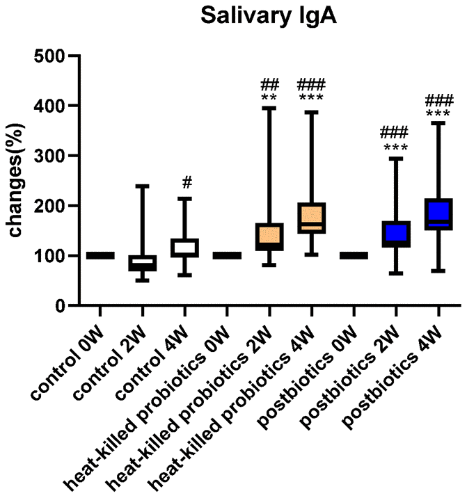
Figure 3. Oral lozenges significantly increased salivary IgA levels. Change in Lactobacillus (%) in participants’ saliva at 0, 2, and 4 weeks with the intake of oral lozenges. Oral lozenges contained postbiotics or heat-killed cells. Participants in the control group consumed placebo lozenges without the postbiotic content (*p < 0.05, **p < 0.01, and ***p < 0.001 compared with the control group; #p < 0.05, ##p < 0.01, and ###p < 0.001 in reference to values at week 0). Data are presented as medians (n = 25 in each group).
NGS detected oral microbiota changes with oral lozenge intake
We used the NGS technique to analyze microbiota changes in saliva samples with oral lozenge intake. Species heatmap (%) demonstrated that L. salivarius significantly increased to 0.03% (compared with the placebo group, p < 0.05) at 4 weeks after consuming postbiotic oral lozenges. Additionally, heat-killed probiotic lozenges significantly increased L. salivarius to 0.06% (compared with the placebo group, p < 0.05; Supplementary Figure 3). The result confirmed our previous findings on plate culturing for quantifying Lactobacillus in saliva samples (Figure 2D).
LEfSe analysis was used to identify the oral microbiota change between before and after oral lozenge intake. Nine differential bacterial taxa significantly increased after the intake of heat-killed probiotic lozenges, including Lactobacillus (Figure 4A). In total, 10 oral bacterial clades significantly increased with the intake of postbiotic lozenges, including Lactobacillus (Figure 4B).
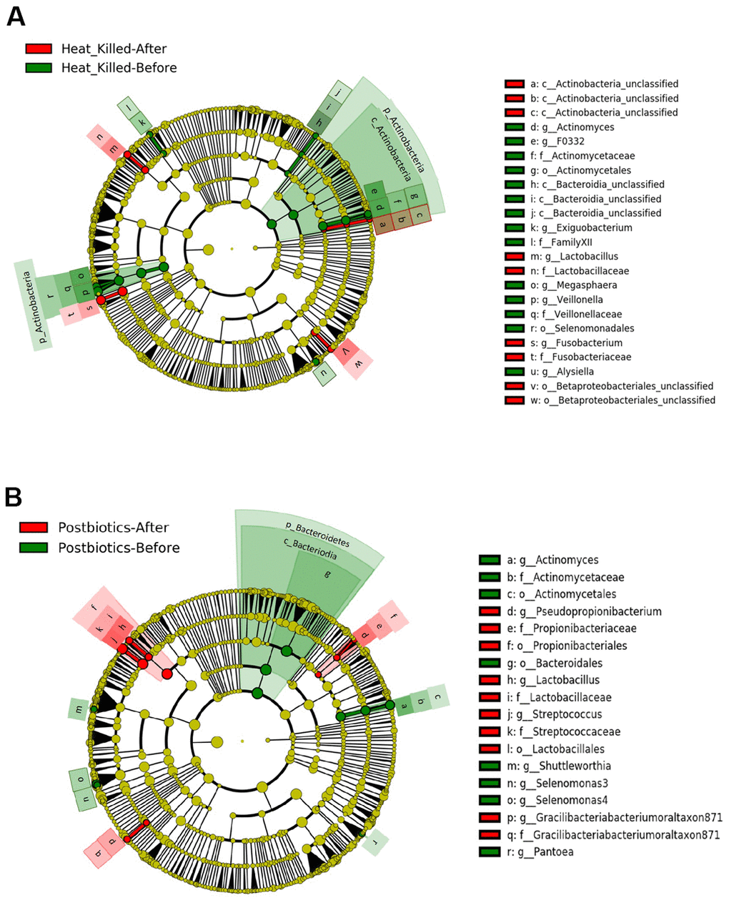
Figure 4. LEfSe analysis of differential oral microbiota before and after 4 weeks of consuming oral lozenge. Comparing changes in oral microbiota with the intake of (A) heat-killed probiotic lozenges and (B) postbiotic lozenges (n = 25 in each group).
We further analyzed statistical alteration in certain oral bacterial strains with the intake of heat-killed probiotic or postbiotic lozenges (Figure 5A–5F). Pathogenic Veillonella spp. (p < 0.01), Actinomyces graevenitzii F0530 (p < 0.05), and Prevotella sp. C561 (p < 0.001) significantly decreased by 4 weeks of treatment with heat-killed probiotic lozenges. However, the postbiotic oral lozenges significantly reduced the growth of Selenomonas 3 spp. (p < 0.01) and Prevotella sp. oral clone FW035 (p < 0.01). L. salivarius significantly increased with the intake of heat-killed probiotic (p < 0.01) and postbiotic (p < 0.001) lozenges.
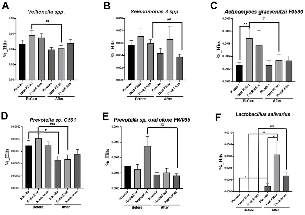
Figure 5. Significant changes in specific oral bacterial strains after consuming heat-killed probiotic or postbiotic oral lozenges. Changes in (A) Veillonella spp., (B) Selenomonas 3 spp., (C) Actinomyces graevenitzii F0530, (D) Prevotella sp. C561, (E) Prevotella sp. oral clone FW035, and (F) L. salivarius after consuming heat-killed probiotic or postbiotic oral lozenges were analyzed through the LEfSe analysis. Comparing changes in oral bacterial concentration (%) in participants’ saliva at 0 (before) or 4 weeks (after) of oral lozenge intake. The oral lozenges contained postbiotics or heat-killed cells. Participants in the control group consumed placebo lozenges without the postbiotic content (*p < 0.05, **p < 0.01, and ***p < 0.001 compared with the control group; #p < 0.05, ##p < 0.01, and ###p < 0.001 in reference to the values at week 0). Two-tailed t-tests was performed to analyze the statistical difference of experimental results. Data are presented as means ± SDs (n = 25 in each group).
Oral lozenges relieved the symptoms of mouth sores, constipation, and gastroesophageal reflux based on health questionnaire analysis
The severity scores for mouth sores or pustule formation decreased to 0.04 ± 0.2 (p < 0.01, compared with the placebo group) and 0.08 ± 0.28 (p < 0.05, compared with the placebo group) after postbiotic lozenge administration for 2 and 4 weeks, respectively (Supplementary Table 1). Furthermore, the heat-killed lozenges relieved the symptoms of mouth sores at weeks 2 (0.23 ± 0.51, p < 0.05, compared with the placebo group) and 4 (0.15 ± 0.37, p < 0.05, compared with the placebo group), respectively. The scores for constipation declined to 0.12 ± 0.33 (p < 0.01) after 4 weeks of postbiotic lozenge intake (Supplementary Table 2). Additionally, 4 weeks after postbiotic oral lozenge intake, the symptoms of gastroesophageal reflux, cold, and drowsiness significantly decreased to 0.12 ± 0.33 (p < 0.05), 0.16 ± 0.37 (p < 0.05), and 0.12 ± 0.33 (p < 0.01), respectively. Moreover, the heat-killed probiotic lozenges diminished the symptoms of constipation, gastroesophageal reflux, cold, and drowsiness at week 4 to 0.15 ± 0.46 (p < 0.05), 0.19 ± 0.4 (p < 0.05), 0.12 ± 0.43 (p < 0.05), and 0.08 ± 0.27 (p < 0.01), respectively.
Discussion
Based on previous research of viable strain-specific approach [25], we selected three of most appropriate strains to generate oral health promoting products of heat-killed probiotic (ET-66 and AP-32), and postbiotic lozenges (LPL28, ET-66, and AP-32) [22, 23]. Several studies have tested the effect of certain live probiotic strains on oral health. For example, probiotic Streptococcus salivarius was reported to reduce severe oral halitosis [26]. However, no study has investigated the role of the postbiotics on regulating oral microbiota and oral immunity. At the beginning of this research, we used in vitro antipathogenic assay to demonstrate that individual postbiotics of AP-32, ET-66, and LPL28 can limit the growth rate of oral pathogenic bacteria S.mutans and periodontal pathogens P. gingivalis BCRC 17689, P. gingivalis BCRC 17688, F. nucleatum subsp. polymorphum, and A. actinomycetemcomitans (Figure 1A). Furthermore, the postbiotic lozenge made from mixed metabolites of AP-32, ET-66, and LPL28 exhibited reliable antibacterial function in vitro (Figure 1B). The heat-killed probiotic lozenges showed better inhibition rate in S. mutans and A. actinomycetemcomitans than postbiotic lozenges. Moreover, the heat-killed probiotic lozenges presented excellent bactericidal ability in oral pathogens, which was in accordance with previous findings on individual heat-killed strains [22]. However, the oral lozenges (made from 3 mixed postbiotics) did not inhibit S. mutans and A. actinomycetemcomitans better than individual ET-66 postbiotic did. This may be because of a lower concentration of functional ingredients in the oral lozenge than in an individual postbiotic (50 mg individual postbiotic versus 50 mg of mixed postbiotics/1 g of lozenge). Besides, previous study revealed viable probiotic lozenges had an excellent inhibition rate (nearly 100%) in five oral pathogenic bacteria S. mutans and P. gingivalis BCRC 17689, P. gingivalis BCRC 17688, F. nucleatum subsp. polymorphum, and A. actinomycetemcomitans [23]. Higher dosage of heat-killed and postbiotic lozenges are presumed to achieve similar pathogenic growth inhibition rate to viable probiotic lozenges. The half maximal inhibitory concentration (IC50) for heat-killed and postbiotic lozenges in limiting oral growth rate should be tested in future.
Next, we validated the antipathogenic ability of heat-killed probiotic or postbiotic lozenges through the detection of changes in the microbial number in saliva.
Compared with the placebo group, the postbiotic and heat-killed lozenge groups exhibited significantly reduced numbers of S. mutans and total bacteria at week 4, but no significantly difference at week 2 (Figure 2A, 2B). S. mutans is the main pathogen involved in the initiation of dental caries and exhibited a positive correlation with periodontitis [27]. In addition, a high ratio of S. mutans DNA was discovered in cardiovascular specimens [28]. Thus, reducing oral S. mutans numbers with postbiotic lozenge intake may prevent dental cavity progression, periodontitis, and cardiovascular diseases. Moreover, Bifidobacterium and Lactobacillus in saliva (cultured on MRS agar plate) revealed that cell numbers increased with the intake of heat-killed probiotic or postbiotic lozenges (Figure 2C, 2D).
Furthermore, postbiotic or heat-killed probiotic lozenges increased IgA concentration in saliva (Figure 3). IgA constitutes 10%–20% of the serum immunoglobulin, second only to IgG. Moreover, IgA present in the mucosal tissues of the oral cavity, digestive tract, and respiratory tract prevents pathogen invasion. Additionally, IgA is present in saliva, tears, and breast milk, particularly in that with high colostrum. No IgA antibody is present in neonatal serum, but newborns obtain IgA secreted from breast milk [29]. Carbohydrate intake may reduce IgA concentration in saliva [30]. Furthermore, salivary IgA acts as the frontline mucosal immune defense against the entry of respiratory pathogens, including severe acute respiratory syndrome coronavirus 2 [31]. Therefore, novel postbiotic or heat-killed probiotic lozenges, which effectively increase salivary IgA concentration, improve bacteriostatic activities, and increase oral populations of beneficial bacteria, may be a potential food product in improving oral health and preventing further infection.
Additionally, we used LEfSe analysis to detect significant changes in oral microbiota after the administration of heat-killed probiotic or postbiotic lozenges (Figure 4). Both heat-killed and postbiotic lozenges showed the ability to significantly increase Lactobacillus in the oral cavity. Furthermore, heatmap results of NGS analysis showed an increase in L. salivarius level (Supplementary Figure 3). Thus, oral lozenges containing postbiotic or heat-killed probiotic cells promoted the growth and colonization of beneficial microorganism in the oral cavity. In addition, the upregulation of L. salivarius in oral microbiota has been reported to promote anticariogenic effects [32, 33]. Moreover, an animal study revealed that L. salivarius subsp. salicinius AP-32 can eradicate Helicobacter pylori infection in addition to improving oral health [34].
Based on previous findings that probiotic strains of AP-32, ET-66 and LPL28 effected oral microbiota [23] and viable strains may improve salivary IgA via up-regulating anti-inflammatory cytokines, IL-10 and TGF-beta [35]. Besides, a previous animal study discovered the mixed viable probiotic strains of Lactobacillus salivarius subsp. salicinius AP-32, L. johnsonii MH-68, L. reuteri GL-104, and Bifidobacterium animalis subsp. lactis CP-9, significantly increased SCFA and MCFA levels. The elevated SCFA and MCFA levels may affect the populations of gut microbiota [36]. The secreted SCFA and MCFA from viable probiotic strains may affect the oral bacterial populations. Some metabolites such as butyrate may stimulate the formation of periodontal/periapical tissues at low or high concentrations [37–39].
However, the detailed mechanism of how three mixed viable probiotic strains (AP-32, ET-66, and LPL28) altered the oral microbiome should be tested in the future. Here, we further discovered that postbiotic would also improve oral microbiota. Nevertheless, the clinical oral health improving function should be tested after stopping consuming postbiotic lozenges. A larger scale clinical analysis of oral microbiota and metabolite profiling for the development of personalized oral therapy in the future [40].
The heat-killed lozenges were efficacious in reducing growth of pathogenic Veillonella spp., A. graevenitzii F0530, and Prevotella sp. C561. Moreover, the postbiotic lozenges reduced the growth of Selenomonas 3 spp. and Prevotella sp. oral clone FW035 (Figure 5). Veillonella spp. has been reported to be associated with halitosis [41] and dental caries [42]. The overgrowth of Prevotella spp. may lead to halitosis [41] and periodontal disease [43]. A. graevenitzii has been discovered to cause pulmonary abscess [44], pneumonia [45], and dental caries [42]. Thus, heat-killed probiotic lozenges might improve the oral smell and oral hygiene by reducing the oral population of Veillonella spp., Prevotella spp., and A. graevenitzii. Additionally, postbiotic oral lozenges may significantly reduce Selenomonas spp. and Prevotella spp., which are associated with halitosis [41]. Moreover, the oral health questionnaires presented that heat-killed and postbiotic lozenges would significantly improve symptoms of ruptured mouth, drool (Supplementary Table 1), constipation, gastroesophageal reflux, cold, drowsiness (Supplementary Table 2). The results of questionnaires at present study are in accordance with previous findings in viable probiotic lozenges. The questionnaires for viable probiotic lozenge present additional improvements in teeth bleeding, sore throat, and stomach pain [23].
Finally, Ishikawa, K. H. et al. demonstrated postbiotics would effectively limit the formation of biofilm formation and growth rate of A. actinomycetemcomitans [21]. At present study, it demonstrated that postbiotic significantly reduced the survival rate of other oral pathogens including S. mutans and periodontal pathogens P. gingivalis BCRC 17689, P. gingivalis BCRC 17688, F. nucleatum subsp. polymorphum. We also measured changes in participants’ salivary IgA and oral microbiota by consuming lozenges of postbiotic AP-32 (L. salivarius subsp. salicinius), ET-66 (L. paracasei), and LPL28 (L. plantarum). The different mixing proportion of three postbiotics effected on oral hygiene should be tested in future.
In conclusion, the postbiotics and heat-killed probiotics have the advantages of preservation and stable viability over viable strains. Here, we found that lozenges of postbiotic AP-32 (L. salivarius subsp. salicinius), ET-66 (L. paracasei), and LPL28 (L. plantarum) and the heat-killed probiotics of AP-32 (L. salivarius subsp. salicinius) and ET-66 (L. paracasei) were beneficial to oral health. Previous study demonstrated that three strains had excellent antimicrobial activity in zone of inhibition test. The present clinical study revealed that postbiotic or heat-killed probiotic lozenges could effectively reduce the number of S. mutans in the oral cavity, increase L. salivarius in oral microbial flora, increase salivary IgA concentration, and decrease oral infections. Furthermore, results from the subjective questionnaire revealed that improved oral health was associated with attenuated intestinal symptoms, relieved constipation, and reduced gastroesophageal reflux, stomach pain, colds, and sense of drowsiness. This study suggested that deactivated probiotic cells and their postbiotics can serve as supporters to optimize the efficacy of oral health supplements. However, further experiments are required.
According to the current manufacturing regulations of cosmetics and cleaning products in various countries, the inactive substances of functional lactic acid bacteria are more suitable for the industrial application. Therefore, this study presented potential food-grade supplementations for promoting oral health and applicable food industrial products in the future.
Materials and Methods
Oral lozenges of heat-killed probiotics and postbiotics
Three probiotic strains known for their antipathogenic against oral pathogens, namely L. salivarius subsp. salicinius AP-32, L. paracasei ET-66, and L. plantarum LPL28, were obtained from Bioflag biotech. Co. Ltd (Tainan, Taiwan). L. salivarius subsp. salicinius AP-32 was isolated from healthy human intestine and deposited in Food Industry Research and Development Institute, Taiwan (ID: BCRC 910437) and in Wuhan university, China (ID: CCTCC-M2011127); L. paracasei ET-66 was isolated from healthy human breast milk and deposited in Food Industry Research and Development Institute, Taiwan (ID: BCRC 910753) and in China General Microbiological Culture Collection Center, Beijing, China (ID: CGMCC-13514). L. plantarum LPL28 was isolated from fermented food Mizo and deposited in Food Industry Research and Development Institute, Taiwan (ID: BCRC 910536) and in China General Microbiological Culture Collection Center, Beijing, China (ID: CGMCC-17954).
We collected postbiotics from these three strains (50 mg of mixed postbiotics/1 g of lozenge) to develop oral lozenges [46]. The detailed procedure for producing postbiotic powder is described as follow: Incubating three probiotic strains AP-32, ET-66 and LPL28 (2 × 10¹¹ colony-formation units [CFUs]/g) in De Man, Rogosa and Sharpe (MRS) media (Difco. Laboratories, Detroit, MI, USA) at 37° C for 48 h to obtain viable probiotics strains. Fermenting mixed probiotics stains of AP-32, ET-66 and LPL28 (the concentration of each probiotic strain was 1 × 10⁹ CFU/mL) with nitrogen sources (skimmed milk and soy bean) and carbohydrate sources (glucose, fructose) at 37° C for 16 hours. Collecting fermented supernatant (postbiotic solution) after centrifugation at 15,000 x g. Pasteurizing fermented product with ultra-high-temperature (UHT) to 135–140° C for 4 seconds. Then spray-drying fermented solution into postbiotic powder. The major nutritional components of postbiotic powder (per 100 g contribution) were crude protein 15.9 g, crude fat 1.9 g, saturated fat 0.21 g, carbohydrate 65.3 g, sugar 3.799 g, glucose 0.553 g, sucrose 0.082 g, maltose 0.667 g, lactose 2.497 g, sodium 3062.7 mg, and calories 341.9 Kcal.
We incubated two probiotic strains L. salivarius subsp. salicinius AP-32 and L. paracasei ET-66 (2 × 10¹¹ colony-formation units [CFUs]/g) in De Man, Rogosa and Sharpe (MRS) media (Difco. Laboratories, Detroit, MI, USA) at 37° C for 48 h to obtain viable probiotics strains [22]. Fermentation and centrifugation procedure was the same as making postbiotic product. Collecting and pasteurizing pellet with ultra-high-temperature (UHT) to 135–140° C for 4 seconds. Freeze-drying the pasteurized pellet for 40 hr, and then obtaining heat-killed probiotic powder. The heat-killed probiotic oral lozenges were composed of 1010 CFUs/g of cells. Furthermore, food-grade D-sorbitol, erythritol, fructooligosaccharides, lactose, magnesium stearate, silica, and sucralose were used to prepare placebo oral lozenges. Furthermore, food-grade D-sorbitol, erythritol, fructooligosaccharides, lactose, magnesium stearate, silica, and sucralose were used to prepare placebo oral lozenges.
Oral pathogenic bacteria
We used tryptic soy broth (TSB; Merck KGaA, Darmstadt, Germany) supplemented with 5% sheep’s blood to cultivate P. gingivalis and F. nucleatum subsp. polymorphum and brain heart infusion (BHI; Merck KGaA, Darmstadt, Germany) broth for culturing A. actinomycetemcomitans. Additionally, TSB was used to cultivate Streptococcus mutans. We incubated pathogens at 37° C (48 h) for subsequent antibacterial tests. S. mutans BCRC 10793T, P. gingivalis BCRC 17689, P. gingivalis BCRC 17688, F. nucleatum subsp. polymorphum BCRC 17679, and A. actinomycetemcomitans BCRC 14405 were obtained from Bioresource Collection and Research Center (BCRC), Hsinchu, Taiwan.
Analyzing bacteriostatic activities
The three probiotic strains were individually cultured in MRS media at 37° C for 20 h. Then, 4.9 mL of supernatants were collected and mixed with oral pathogenic bacteria (106 CFUs/0.1 mL) after which the mixed solution was incubated at 37° C for 48 h. Subsequently, the CFUs of pathogenic bacteria in each tube were calculated. Furthermore, the CFUs of oral pathogens were compared with the control media, which contained pathogens without postbiotic treatment.
The bacteriostatic activities of postbiotic and heat-killed probiotic lozenges were tested according to the same protocol. The experimental lozenges were dissolved in either a TSB or BHI medium at 0.1 g/mL concentration, and then, oral pathogens (106 CFU) were introduced into the lozenge solutions and coincubated at 37° C for 2 (S. mutans) or 3 (P. gingivalis, F. nucleatum subsp. polymorphum, and A. actinomycetemcomitans) days. Furthermore, the CFUs of pathogenic bacteria in each tube were calculated. We measured the survival rates of the oral pathogens by using the following formula: CFUexperimental group/CFUcontrol media (%). The inhibition rates of the oral pathogens were determined using the following formula: 1 − survival rate (%). The metabolites of L. rhamnosus GG (LGG) purchased from Chr. Hansen, Hoersholm, Denmark were tested as positive control.
Participants
In total, 75 participants (on-smokers, free from systemic diseases) between 20 and 40 years of age and with S. mutans >105 CFUs/mL in their saliva samples were recruited. Their average age was 26.29 ± 5.59 years. The initial amount of S. mutans in their saliva (CFUs/mL) was 4.25* 106 ± 2.90* 106, and their plaque weight was 0.37 ± 0.16 g. All clinical tests were performed according to the guidelines of the Ministry of Health and Welfare, Taiwan (Health Food Evaluation No. 88037803). All participants were randomly and blindly assigned to three groups: placebo, heat-killed probiotics, and postbiotics (25 participants in each group). Participants were asked to clean their oral cavity and then consume three oral lozenges (3 g) every day for 4 weeks [47]. We collected and measured oral microbiota, IgA levels, and oral pathogens in 2-mL saliva samples at weeks 0, 2, and 4. Additionally, total plaque and oral health questionnaire were analyzed at weeks 0, 2, and 4. The protocols for evaluating the uptake of postbiotic products, colleting human saliva samples, and administering subjective questionnaires were approved by the Institutional Review Board of Chung Shan Medical University, Taiwan (CS19052).
Analysis of the populations of Lactobacillus, Bifidobacterium, S. mutans, and total aerobic bacteria in the oral cavity
For analyzing the populations of Lactobacillus, Bifidobacterium, total aerobic bacteria, and S. mutans in the oral cavity, 100-μL saliva samples were cultured on MRS with 0.05% cysteine agar, plate count agar (PCA; Merck KGaA, Darmstadt, Germany), and mitis salivarius-bacitracin (MSB Agar) (Merck KGaA, Darmstadt, Germany) in triplicate, and CFUs on each plate were calculated. The change rates of oral pathogen were determined using the following formula: (CFUsweek 2 or 4 − CFUsweek 0)/CFUsweek 0 (%).
Next-generation sequencing analysis of oral microbiota
Changes in oral microbiota were measured using the next-generation sequencing (NGS) technique. Microbial DNA was extracted from the saliva samples and sent to Genomics Co. Ltd. for NGS analysis. In brief, commercial specific primers (Genomics Co. Ltd., Taiwan) were used to amplify the amplicon DNA segments (16S rRNA and 16S V3–V4) by using the polymerase chain reaction (PCR) technique (Phusion High-Fidelity PCR Master Mix, New England Biolabs, USA). The PCR products of 400–450 bp were purified using the Qiagen Gel Extraction kit (Qiagen, Germany). Then, the TruSeq DNA PCR-free sample preparation kit (Illumina, USA) was used to generate sequencing libraries with provided index codes. The Qubit 2.0 Fluorometer (Thermo Scientific, USA) and Agilent Bioanalyzer 2100 system (Agilent Technologies, Inc., USA) were applied to confirm the library quality. Finally, the Illumina HiSeq 2500 platform was used to sequence and analyze the DNA library. QIIME software, version 1.7.0, was used to analyze NGS raw data [48]. The sizes of each single taxon of the groups were further analyzed through linear regression plots and linear discriminant analysis effect size (LEfSe) analysis (https://huttenhower.sph.harvard.edu/galaxy/) and the analysis protocol was followed the instruction in https://twbattaglia.gitbooks.io/introduction-to-qiime/content/lefse.html.
Measuring IgA and plaque
The human IgA enzyme-linked immunosorbent assay (ELISA) kit (Invitrogen, Lot: 218315-003) was used to measure IgA concentrations of saliva samples in triplicate. The IgA concentration was analyzed at an optical density of 450–570 nm by using the ELISA reader. Plaque in the mouth was collected using a swab. Then, the dehydrated plaque was weighed. The weight of plague (g) = total weight of samples with sample tubes (g) – sample tubes (g).
Questionnaire of dental problems and gastrointestinal symptoms
Self-report questionnaires were used to evaluate common dental symptoms and a gastrointestinal symptom [23]. All participants completed the questionnaire at weeks 0, 2, and 4 after the intervention. Participants could give the following responses: 0 = no symptom; 1 = mild; 2 = medium; 3 = serious.
Statistics
GraphPad Prism software (San Diego, CA, USA) was applied to perform statistical analysis of collected data. Data of bacterial colonies in oral and salivary IgA are presented as medians. Each test was performed triplicate. The rest of the data are presented as means ± standard deviations (SDs) or means. Two-tailed t-tests were performed to analyze the statistical differences in experimental results. Statistical difference was indicated by p < 0.05. A significant statistical difference was observed in the treatment data of the experimental groups compared with their pretreatment data at week 0 (# p < 0.05) and compared with the placebo group data at week 4 (* p < 0.05).
Author Contributions
Conceptualization, Chiao-Wen Lin, Yi-Tzu Chen, Shun-Fa Yang and Hsieh-Hsun Ho; Methodology, Yi-Wei Kuo, Yao-Tsung Yeh, Ching-Wei Chen, Yu-Fen Huang, and Chen-Hung Hsu; Investigation: Wen-Yang Lin; Formal analysis: Yi-Wei Kuo; Data acquisition and classification: Jia-Hung Lin, Chi-Huei Lin, Cheng-Ruei Liu and Jui-Fen Chen; Data Visualization, Wen-Yang Lin; Writing - original draft preparation, Wen-Yang Lin; Writing - review and editing: Chiao-Wen Lin, Hsieh-Hsun Ho, Yi-Wei Kuo, Wen-Yang Lin, Pei-Shan Hsieh and Shun-Fa Yang.
Conflicts of Interest
The authors declare that they have no conflicts of interest.
Funding
Bioflag Biotech Co., Ltd provided support in the form of salaries for authors, but did not have any additional role in the study design, data collection and analysis, decision to publish, or preparation of the manuscript.
References
- 1. Wu RQ, Zhang DF, Tu E, Chen QM, Chen W. The mucosal immune system in the oral cavity-an orchestra of T cell diversity. Int J Oral Sci. 2014; 6:125–32. https://doi.org/10.1038/ijos.2014.48 [PubMed]
- 2. McLean JS. Advancements toward a systems level understanding of the human oral microbiome. Front Cell Infect Microbiol. 2014; 4:98. https://doi.org/10.3389/fcimb.2014.00098 [PubMed]
- 3. Perera M, Al-Hebshi NN, Speicher DJ, Perera I, Johnson NW. Emerging role of bacteria in oral carcinogenesis: a review with special reference to perio-pathogenic bacteria. J Oral Microbiol. 2016; 8:32762. https://doi.org/10.3402/jom.v8.32762 [PubMed]
- 4. Sharma N, Bhatia S, Sodhi AS, Batra N. Oral microbiome and health. AIMS Microbiol. 2018; 4:42–66. https://doi.org/10.3934/microbiol.2018.1.42 [PubMed]
- 5. Jia G, Zhi A, Lai PF, Wang G, Xia Y, Xiong Z, Zhang H, Che N, Ai L. The oral microbiota - a mechanistic role for systemic diseases. Br Dent J. 2018; 224:447–55. https://doi.org/10.1038/sj.bdj.2018.217 [PubMed]
- 6. Kilian M, Chapple IL, Hannig M, Marsh PD, Meuric V, Pedersen AM, Tonetti MS, Wade WG, Zaura E. The oral microbiome - an update for oral healthcare professionals. Br Dent J. 2016; 221:657–66. https://doi.org/10.1038/sj.bdj.2016.865 [PubMed]
- 7. Dewhirst FE, Chen T, Izard J, Paster BJ, Tanner AC, Yu WH, Lakshmanan A, Wade WG. The human oral microbiome. J Bacteriol. 2010; 192:5002–17. https://doi.org/10.1128/JB.00542-10 [PubMed]
- 8. Nath SG, Raveendran R. Microbial dysbiosis in periodontitis. J Indian Soc Periodontol. 2013; 17:543–5. https://doi.org/10.4103/0972-124X.118334 [PubMed]
- 9. Jepsen K, Jepsen S. Antibiotics/antimicrobials: systemic and local administration in the therapy of mild to moderately advanced periodontitis. Periodontol 2000. 2016; 71:82–112. https://doi.org/10.1111/prd.12121 [PubMed]
- 10. Bolstad AI, Jensen HB, Bakken V. Taxonomy, biology, and periodontal aspects of Fusobacterium nucleatum. Clin Microbiol Rev. 1996; 9:55–71. https://doi.org/10.1128/CMR.9.1.55 [PubMed]
- 11. Nazir MA. Prevalence of periodontal disease, its association with systemic diseases and prevention. Int J Health Sci (Qassim). 2017; 11:72–80. [PubMed]
- 12. Blaizot A, Vergnes JN, Nuwwareh S, Amar J, Sixou M. Periodontal diseases and cardiovascular events: meta-analysis of observational studies. Int Dent J. 2009; 59:197–209. [PubMed]
- 13. Petersen PE, Ogawa H. Strengthening the prevention of periodontal disease: the WHO approach. J Periodontol. 2005; 76:2187–93. https://doi.org/10.1902/jop.2005.76.12.2187 [PubMed]
- 14. Morales A, Gandolfo A, Bravo J, Carvajal P, Silva N, Godoy C, Garcia-Sesnich J, Hoare A, Diaz P, Gamonal J. Microbiological and clinical effects of probiotics and antibiotics on nonsurgical treatment of chronic periodontitis: a randomized placebo- controlled trial with 9-month follow-up. J Appl Oral Sci. 2018; 26:e20170075. https://doi.org/10.1590/1678-7757-2017-0075 [PubMed]
- 15. de Vos P, Faas MM, Spasojevic M, Sikkema J. Encapsulation for preservation of functionality and targeted delivery of bioactive food components. Int Dairy J. 2010; 20:292–302. https://doi.org/10.1016/j.idairyj.2009.11.008
- 16. Yamada M, Takahashi N, Matsuda Y, Sato K, Yokoji M, Sulijaya B, Maekawa T, Ushiki T, Mikami Y, Hayatsu M, Mizutani Y, Kishino S, Ogawa J, et al. A bacterial metabolite ameliorates periodontal pathogen-induced gingival epithelial barrier disruption via GPR40 signaling. Sci Rep. 2018; 8:9008. https://doi.org/10.1038/s41598-018-27408-y [PubMed]
- 17. Salminen S, Collado MC, Endo A, Hill C, Lebeer S, Quigley EM, Sanders ME, Shamir R, Swann JR, Szajewska H, Vinderola G. The International Scientific Association of Probiotics and Prebiotics (ISAPP) consensus statement on the definition and scope of postbiotics. Nat Rev Gastroenterol Hepatol. 2021; 18:649–67. https://doi.org/10.1038/s41575-021-00440-6 [PubMed]
- 18. Wegh CA, Geerlings SY, Knol J, Roeselers G, Belzer C. Postbiotics and Their Potential Applications in Early Life Nutrition and Beyond. Int J Mol Sci. 2019; 20:4673. https://doi.org/10.3390/ijms20194673 [PubMed]
- 19. Ryan PM, Ross RP, Fitzgerald GF, Caplice NM, Stanton C. Sugar-coated: exopolysaccharide producing lactic acid bacteria for food and human health applications. Food Funct. 2015; 6:679–93. https://doi.org/10.1039/C4FO00529E [PubMed]
- 20. Rad AH, Abbasi A, Kafil HS, Ganbarov K. Potential Pharmaceutical and Food Applications of Postbiotics: A Review. Curr Pharm Biotechnol. 2020; 21:1576–87. https://doi.org/10.2174/1389201021666200516154833 [PubMed]
- 21. Ishikawa KH, Bueno MR, Kawamoto D, Simionato MR, Mayer MP. Lactobacilli postbiotics reduce biofilm formation and alter transcription of virulence genes of Aggregatibacter actinomycetemcomitans. Mol Oral Microbiol. 2021; 36:92–102. https://doi.org/10.1111/omi.12330 [PubMed]
- 22. Chen YT, Hsieh PS, Ho HH, Hsieh SH, Kuo YW, Yang SF, Lin CW. Antibacterial activity of viable and heat-killed probiotic strains against oral pathogens. Lett Appl Microbiol. 2020; 70:310–17. https://doi.org/10.1111/lam.13275 [PubMed]
- 23. Lin CW, Chen YT, Ho HH, Hsieh PS, Kuo YW, Lin JH, Liu CR, Huang YF, Chen CW, Hsu CH, Lin WY, Yang SF. Lozenges with probiotic strains enhance oral immune response and health. Oral Dis. 2021. [Epub ahead of print]. https://doi.org/10.1111/odi.13854 [PubMed]
- 24. Atlas RM. Handbook of microbiological media. CRC press. 2004. https://doi.org/10.1201/9781420039726
- 25. Bubnov RV, Babenko LP, Lazarenko LM, Mokrozub VV, Spivak MY. Specific properties of probiotic strains: relevance and benefits for the host. EPMA J. 2018; 9:205–23. https://doi.org/10.1007/s13167-018-0132-z [PubMed]
- 26. Burton JP, Chilcott CN, Moore CJ, Speiser G, Tagg JR. A preliminary study of the effect of probiotic Streptococcus salivarius K12 on oral malodour parameters. J Appl Microbiol. 2006; 100:754–64. https://doi.org/10.1111/j.1365-2672.2006.02837.x [PubMed]
- 27. Dani S, Prabhu A, Chaitra KR, Desai NC, Patil SR, Rajeev R. Assessment of Streptococcus mutans in healthy versus gingivitis and chronic periodontitis: A clinico-microbiological study. Contemp Clin Dent. 2016; 7:529–34. https://doi.org/10.4103/0976-237X.194114 [PubMed]
- 28. Nakano K, Nomura R, Ooshima T. Streptococcus mutans and cardiovascular diseases. Jpn Dent Sci Rev. 2008; 44:29–37. https://doi.org/10.1016/j.jdsr.2007.09.001
- 29. Pabst O. New concepts in the generation and functions of IgA. Nat Rev Immunol. 2012; 12:821–32. https://doi.org/10.1038/nri3322 [PubMed]
- 30. Bishop NC, Blannin AK, Armstrong E, Rickman M, Gleeson M. Carbohydrate and fluid intake affect the saliva flow rate and IgA response to cycling. Med Sci Sports Exerc. 2000; 32:2046–51. https://doi.org/10.1097/00005768-200012000-00013 [PubMed]
- 31. Chao YX, Rötzschke O, Tan EK. The role of IgA in COVID-19. Brain Behav Immun. 2020; 87:182–3. https://doi.org/10.1016/j.bbi.2020.05.057 [PubMed]
- 32. Nishihara T, Suzuki N, Yoneda M, Hirofuji T. Effects of Lactobacillus salivarius-containing tablets on caries risk factors: a randomized open-label clinical trial. BMC Oral Health. 2014; 14:110. https://doi.org/10.1186/1472-6831-14-110 [PubMed]
- 33. Krzyściak W, Kościelniak D, Papież M, Vyhouskaya P, Zagórska-Świeży K, Kołodziej I, Bystrowska B, Jurczak A. Effect of a Lactobacillus Salivarius Probiotic on a Double-Species Streptococcus Mutans and Candida Albicans Caries Biofilm. Nutrients. 2017; 9:1242. https://doi.org/10.3390/nu9111242 [PubMed]
- 34. Hsieh PS, Tsai YC, Chen YC, Teh SF, Ou CM, King VA. Eradication of Helicobacter pylori infection by the probiotic strains Lactobacillus johnsonii MH-68 and L. salivarius ssp. salicinius AP-32. Helicobacter. 2012; 17:466–77. https://doi.org/10.1111/j.1523-5378.2012.00992.x [PubMed]
- 35. Lin WY, Kuo YW, Chen CW, Huang YF, Hsu CH, Lin JH, Liu CR, Chen JF, Hsia KC, Ho HH. Viable and Heat-Killed Probiotic Strains Improve Oral Immunity by Elevating the IgA Concentration in the Oral Mucosa. Curr Microbiol. 2021; 78:3541–9. https://doi.org/10.1007/s00284-021-02569-8 [PubMed]
- 36. Hsieh PS, Ho HH, Tsao SP, Hsieh SH, Lin WY, Chen JF, Kuo YW, Tsai SY, Huang HY. Multi-strain probiotic supplement attenuates streptozotocin-induced type-2 diabetes by reducing inflammation and β-cell death in rats. PLoS One. 2021; 16:e0251646. https://doi.org/10.1371/journal.pone.0251646 [PubMed]
- 37. Guan X, Li W, Meng H. A double-edged sword: role of butyrate in the oral cavity and the gut. Mol Oral Microbiol. 2021; 36:121–31. https://doi.org/10.1111/omi.12322 [PubMed]
- 38. Chang MC, Wang TM, Chien HH, Pan YH, Tsai YL, Jeng PY, Lin LD, Jeng JH. Effect of butyrate, a bacterial by-product, on the viability and ICAM-1 expression/production of human vascular endothelial cells: role in infectious pulpal/periapical diseases. Int Endod J. 2022; 55:38–53. https://doi.org/10.1111/iej.13614 [PubMed]
- 39. Chang MC, Chen YJ, Lian YC, Chang BE, Huang CC, Huang WL, Pan YH, Jeng JH. Butyrate Stimulates Histone H3 Acetylation, 8-Isoprostane Production, RANKL Expression, and Regulated Osteoprotegerin Expression/Secretion in MG-63 Osteoblastic Cells. Int J Mol Sci. 2018; 19:4071. https://doi.org/10.3390/ijms19124071 [PubMed]
- 40. Singh TP, Natraj BH. Next-generation probiotics: a promising approach towards designing personalized medicine. Crit Rev Microbiol. 2021; 47:479–98. https://doi.org/10.1080/1040841X.2021.1902940 [PubMed]
- 41. Hampelska K, Jaworska MM, Babalska ZŁ, Karpiński TM. The Role of Oral Microbiota in Intra-Oral Halitosis. J Clin Med. 2020; 9:2484. https://doi.org/10.3390/jcm9082484 [PubMed]
- 42. Jiang S, Gao X, Jin L, Lo EC. Salivary Microbiome Diversity in Caries-Free and Caries-Affected Children. Int J Mol Sci. 2016; 17:1978. https://doi.org/10.3390/ijms17121978 [PubMed]
- 43. Nadkarni MA, Browne GV, Chhour KL, Byun R, Nguyen KA, Chapple CC, Jacques NA, Hunter N. Pattern of distribution of Prevotella species/phylotypes associated with healthy gingiva and periodontal disease. Eur J Clin Microbiol Infect Dis. 2012; 31:2989–99. https://doi.org/10.1007/s10096-012-1651-5 [PubMed]
- 44. Gliga S, Devaux M, Gosset Woimant M, Mompoint D, Perronne C, Davido B. Actinomyces graevenitzii pulmonary abscess mimicking tuberculosis in a healthy young man. Can Respir J. 2014; 21:e75–7. https://doi.org/10.1155/2014/841480 [PubMed]
- 45. Fujita Y, Iikura M, Horio Y, Ohkusu K, Kobayashi N. Pulmonary Actinomyces graevenitzii infection presenting as organizing pneumonia diagnosed by PCR analysis. J Med Microbiol. 2012; 61:1156–8. https://doi.org/10.1099/jmm.0.040394-0 [PubMed]
- 46. Stašková A, Sondorová M, Nemcová R, Kačírová J, Maďar M. Antimicrobial and Antibiofilm Activity of the Probiotic Strain Streptococcus salivarius K12 against Oral Potential Pathogens. Antibiotics (Basel). 2021; 10:793. https://doi.org/10.3390/antibiotics10070793 [PubMed]
- 47. Sutula J, Coulthwaite LA, Thomas LV, Verran J. The effect of a commercial probiotic drink containing Lactobacillus casei strain Shirota on oral health in healthy dentate people. Microb Ecol Health Dis. 2013; 24. https://doi.org/10.3402/mehd.v24i0.21003 [PubMed]
- 48. Caporaso JG, Kuczynski J, Stombaugh J, Bittinger K, Bushman FD, Costello EK, Fierer N, Peña AG, Goodrich JK, Gordon JI, Huttley GA, Kelley ST, Knights D, et al. QIIME allows analysis of high-throughput community sequencing data. Nat Methods. 2010; 7:335–6. https://doi.org/10.1038/nmeth.f.303 [PubMed]