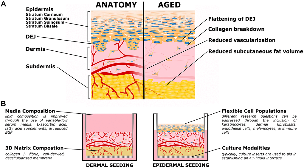
Figure 1.Organotypic models of skin aging. (A) Simplified skin anatomy and aging phenotypes. Skin can be separated into epidermal, dermal, and hypodermal layers. The epidermis is composed of Stratum Basale, Spinosum, Granulosum, and Corneum, composed of increasingly differentiated epidermal cells. The dermal-epidermal junction (DEJ) connects the basement membrane of the Stratum Basale to the upper (papillary) dermis, and is characterized by small dermal extensions (or papilla) into the epidermis. The DEJ flattens with age. The dermis is a collagen rich tissue supported by dermal fibroblasts. The subdermis (or hypodermis) is an important adipose compartment that contributes to overall metabolic function; this tends to thin with age. Both the dermis and subdermis are highly vascularized, important for thermal regulation; in age vascularization is reduced. The above schematic is simplified to focus on the level of current organotypic models, nerves, melanocytes, immune cells, and other components of in vivo skin are not pictured. (B) Organotypic skin models, also referred to as Human Skin Equivalents (HSE), typically consist of a dermal/subdermal culture grown on a permeable culture support (left), followed by seeding and differentiation of epidermis at the air-liquid interface (ALI). Benefits of this style is the accessibility of the culture format, ready customization of the specific cell populations (both immortalized or primary, patient specific, or transgenic disease models), and customization of the matrix and media formulations.