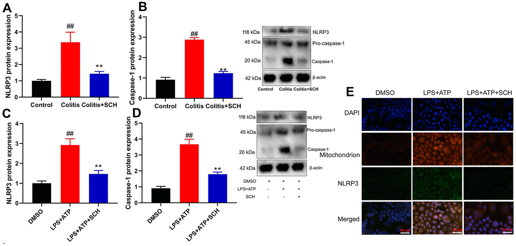
Figure 3.Schisandrin B suppressed NLRP3 inflammasone in vivo and vitro model of colitis. (A, B) the protein expression of NLRP3 and caspase-1 in mouse colon tissue; (C, D) the protein expression of NLRP3 and caspase-1 in intestinal epithelial cells induced by LPS + ATP; (E) the protein expression of NLRP3 and caspase-1 in intestinal epithelial cells detected by cell immunofluorescence. ##P<0.01 vs control group; **P<0.01 vs DSS- induced colitis group. Control: blank control group; Colitis: DSS- induced colitis group; Colitis+SCH: DSS- induced colitis mice with Schisandrin. ##P<0.01 vs MDSO group; **P<0.01 vs LPS+ATP induced intestinal epithelial cells group. MDSO: blank control group; LPS+ATP: intestinal epithelial cells with LPS+ATP group; LPS+ATP +SCH: intestinal epithelial cells induced by LPS+ATP with Schisandrin. SCH, Schisandrin B. Data were expressed as mean ± SEM.