


Introduction
Currently, Parkinson's disease (PD) is the second most common neurodegenerative disease[1, 2]. It affects over seven million people globally [2]. PD is characterized by a progressive loss of dopaminergic (DA) neurons in the midbrain [3–6]. Sustained and unsolved oxidative stress could be one primary cause of DA neuronal cell death. It is also known as the primary pathogenesis mechanism of PD [7].
Disruption of mitochondrial respiratory chain in DA neurons would lead to robust radical oxidative species (ROS) production and oxidative injury. This will then cause calcium overload, lipid peroxidation, DNA breaks, and protein aggregation [7–9]. These events together lead to DA neuronal cell death [7]. The mitochondrial respiratory chain complex inhibitor 1-methyl-4-phenyl-1,2,3,4-tetrahydropyridine (MPTP) has been applied to establish animal PD models [7]. In the brain of primates, MPTP is converted into MPP+ (1-methyl-4-phenylpyridinium) by monoamine oxidase B (MAO-B), causing parkinsonism by killing DA neurons in substantia nigra [7]. Additionally, as a commonly-utilized cellular model of PD, MPP+ is added to the cultured neurons and neuronal cells [10–13].
Sphingosine kinase 1 (SphK1) phosphorylates sphingosine to sphingosine-1-phosphate (S1P) to promote cell survival [14–16]. Conversely, SphK1 inhibition, silencing, or knockout would induce sphingosine/ceramide accumulation and S1P depletion that lead to cell death [16]. SphK1 is expressed in neurons and progenitor cells, and is enriched in cellular membranes of endocytosis, synapses, and mitochondria [17]. Neuronal SphK1 expression is decreased in Alzheimer’s disease (AD) brain, indicating its potential role in the pathogenesis of AD. Upregulation of neuronal SphK1 could promote microglia-induced phagocytosis of aggregated amyloid β (Aβ) and improve cognitive deficits [18]. SphK1-S1P signaling is essential in regulating sensory ganglia development and survival of neurons and progenitor cells [19]. In motor neurons, SphK1 functions in Nrf2 signaling cascade to regulate neuropeptide biogenesis and secretion [19].
Studies have shown that MPP+ serve as a main contributor of subsequent neuronal cell apoptosis as it downregulated SphK1 expression and inhibited its activity [20, 21]. Conversely, SphK1 activation by exogenously adding S1P protected neuronal cells from MPP+-induced cell apoptosis [22]. Therefore, restoring SphK1expression/activity could protect neuronal cells from MPP+ [20, 21].
microRNAs (miRNAs) is a large family of small non-coding RNA molecules containing ~22 nucleotides [23, 24]. miRNAs are capable of silencing targeted genes through a post-transcriptional mechanism [23, 24]. Specifically, miRNAs function via base-pairing with the complementary sequences in the 3’-untranslated region (UTR) of targeted mRNAs/genes. This will cause the degradation and/or translation inhibition of targeted genes [23, 24]. Several SphK1-targeting miRNAs have been identified, and many of them are in cancer cells [25–27]. Here we identifiedmicroRNA-6862 (miR-6862) as a novel SphK1-targeting miRNA. The current study is to determine whether miR-6862 would affect SphK1 expression in neuronal cells, and if so, whether miR-6862 inhibition would protect neuronal cells from MPP+-induced damage.
Results
miR-6862 directly binds to and silences SphK1 in SH-SY5Y neuronal cells
First, the microRNA database TargetScan (V7.2) [28] was consulted to identify miRNAs that can putatively bind to SphK1 3’-UTR [28]. A total of 144 different miRNAs complementary pairing with SphK1 3’-UTR were retrieved,. Eight (8) of them have a context++ score less than -0.5 (Table 1) and context++ score percentile of over 95% (Table 1). These parameters indicated a high percentage of direct binding between these proposed miRNAs and SphK1 3’-UTR [28]. Next, each of the eight miRNA mimics (500 nM for 48h) was individually transfected to SH-SY5Y neuronal cells. Among which microRNA-6862-3p (miR-6862) resulted in the most significant SphK1 mRNA reduction. Thereafter, miRbase (v21.0) and miRDB databases showed that there were over 250 predicted gene targets of miR-6862. Among which, SphK1 is the top 10% most possible targets.
Table 1. miRNAs complementary pairing with SphK1 3’-UTR with context++ score less than -0.5.
| No. | miRNA | Position | Seed match | Context++ score | context++ score percentage |
| 1 | hsa-miR-3677-3p | 235-242 | 8mer | -0.78 | 99 |
| 2 | hsa-miR-6862-3p | 113-120 | 8mer | -0.64 | 99 |
| 3 | hsa-miR-6716-5p | 262-269 | 8mer | -0.61 | 99 |
| 4 | hsa-miR-6784-3p | 113-120 | 8mer | -0.6 | 99 |
| 5 | hsa-miR-4651 | 231-238 | 8mer | -0.59 | 98 |
| 6 | hsa-miR-608 | 231-238 | 8mer | -0.56 | 98 |
| 7 | hsa-miR-5004-3p | 246-253 | 8mer | -0.52 | 99 |
| 8 | hsa-miR-4505 | 266-272 | 7mer-m8 | -0.5 | 99 |
miR-6862 putatively targets the SphK1 3’-UTR (at position 113-120, Figure 1A). The context score percentage of miR-6862-SphK1 3’-UTR binding is 99%. The context++ score is-0.64 (TargetScan V7.2 [28], Figure 1A). Figure 1B demonstrated that miR-6862 fluorescence mainly located in the cytoplasm of SH-SY5Y neuronal cells. Some was in cell nuclei (Figure 1B). Furthermore, RNA-Pull down assay [28] results, Figure 1C, demonstrated that the biotinylated-miR-6862 directly bound to SphK1 mRNA in SH-SY5Y cells (Figure 1C). These results suggested a direct binding between miR-6862 and SphK1 mRNA in neuronal cells.
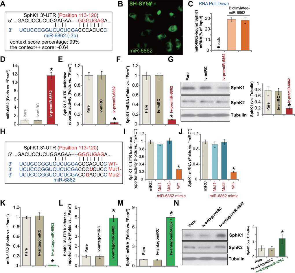
Figure 1. miR-6862 directly binds to and silences SphK1 in SH-SY5Y neuronal cells. miRNA-6862 putatively targets SphK1 mRNA 3’-UTR (untranslated region, at position of 113-120) (A). miRNA-6862 (fluorescence-tagged) locates in the cytoplasm of SH-SY5Y cells (B). RNA pull down showed a directing binding between biotinylated-miR-6862 and SphK1 mRNA in SH-SY5Y cells (C); Stable SH-SY5Y cells with the lentiviral construct encoding the premiR-6862 sequence (lv-premiR-6862) or the nonsense miRNA sequence (lv-miRC) were established, expression of listed genes (mRNA and protein) was shown (D, F, G). The relative SphK1 3’-UTR luciferase reporter activity was tested as well (E). SH-SY5Y cells were transfected with 500 nM of wile-type (“WT-“) or the mutant (“Mut1-”/“Mut2-”) miRNA-6862 mimics (sequences were listed in H), control cells were transfected with nonsense control miRNA (“miRC”), after 48h the relative SphK1 3’-UTR luciferase reporter activity (I) and SphK1 mRNA expression (J) were tested. Stable SH-SY5Y cells with the lentiviral construct encoding the anti-sense of premiR-6862 (lv-antagomiR-6862) or the anti-sense control sequence (lv-antagomiRC) were established, expression of listed genes was shown (K–N). The relative SphK1 3’-UTR luciferase reporter activity was tested as well (L). “Pare” stands for the parental control cells (same for all Figures). Data were presented as mean ± standard deviation (SD, n=5). * P < 0.05 vs. “lv-miRC”/“miRC”/“lv-antagomiRC” cells. Experiments in this figure were repeated five times with similar results obtained.
To test whether miR-6862 can affect SphK1 expression, a lentiviral construct encoding the premiR-6862 sequence (see Table 2) was established. The construct, lv-premiR-6862, was transduced to SH-SY5Y neuronal cells. Via puromycin-mediated selection stable cells were established. The qPCR assay results in Figure 1D demonstrated that the mature miR-6862 expression increased over ten folds (vs. parental control cells) in lv-premiR-6862-expressing SH-SY5Y cells. Subsequently, SphK1 3’-UTR luciferase reporter activity decreased over 90% (Figure 1E). SphK1 mRNA expression was dramatically downregulated as well (Figure 1F). Testing SphK1 protein expression by Western blotting confirmed SphK1 protein downregulation in lv-premiR-6862-expressing SH-SY5Y cells (Figure 1G). SphK2 protein expression, however, was unchanged (Figure 1G). The lentiviral construct encoding nonsense miRNA sequence, lv-miRC, did not change the expression of miR-6862 and SphK1 in SH-SY5Y neuronal cells (Figure 1D–1G).
Table 2. Sequences in this study.
| Genes | Forward sequence (5’-3’) | Reverse sequence (5’-3’) | |||||||||||||||||||||||||||||||||||||||||||||||||||||||||||||||||||||||||||||||||||||||||||||||||
| miR-6862 | CGGGCATGCTGGGAGAGAC | GAACATGTCTGCGTATCTC | |||||||||||||||||||||||||||||||||||||||||||||||||||||||||||||||||||||||||||||||||||||||||||||||||
| U6 RNA | CTCGCTTCGGCAGCACAT | TTTGCGTGTCATCCTTGCG | |||||||||||||||||||||||||||||||||||||||||||||||||||||||||||||||||||||||||||||||||||||||||||||||||
| SphK1 | GCTGGCAGCTTCCTTGAACCAT | GTGTGCAGAGACAGCAGGTTCA | |||||||||||||||||||||||||||||||||||||||||||||||||||||||||||||||||||||||||||||||||||||||||||||||||
| GAPDH | GTCTCCTCTGACTTCAACAGCG | ACCACCCTGTTGCTGTAGCCAA | |||||||||||||||||||||||||||||||||||||||||||||||||||||||||||||||||||||||||||||||||||||||||||||||||
| Primers for qPCR assay. | |||||||||||||||||||||||||||||||||||||||||||||||||||||||||||||||||||||||||||||||||||||||||||||||||||
| miR-6862 precursor sequence: | |||||||||||||||||||||||||||||||||||||||||||||||||||||||||||||||||||||||||||||||||||||||||||||||||||
| CGAAGCGGGCAUGCUGGGAGAGACUUUGUGAUUUGUCUCCAAAGCCUCACCCAGCUCUCUGGCCCUCUAG | |||||||||||||||||||||||||||||||||||||||||||||||||||||||||||||||||||||||||||||||||||||||||||||||||||
| Mature miR-6862 sequence: | |||||||||||||||||||||||||||||||||||||||||||||||||||||||||||||||||||||||||||||||||||||||||||||||||||
| CGGGCAUGCUGGGAGAGACUUU. | |||||||||||||||||||||||||||||||||||||||||||||||||||||||||||||||||||||||||||||||||||||||||||||||||||
To further verify a direct binding between miR-6862 and SphK1 mRNA, we created two mutant miR-6862 mimics containing mutations at the binding sites to SphK1 3’-UTR (Figure 1H). The two were named as “Mut1-” and “Mut2-” (Figure 1H). The wild-type (WT-) and the two mutant miR-6862 mimics were individually transfected in SH-SY5Y cells (500 nM for 48h). As shown, transfection of the WT-miR-6862 resulted in robust decreases in SphK1 3’-UTR luciferase reporter activity (Figure 1I) and SphK1 mRNA expression (Figure 1J). Contrarily, the two mutants, “Mut1-” and “Mut2-”, were completely ineffective (Figure 1I, 1J). These results implied that miR-6862 directly binds to and silences SphK1 in SH-SY5Y cells.
We further hypothesized that miR-6862 inhibition could increase SphK1 expression. Therefore, a lentiviral construct encoding premiR-6862 anti-sense (lv-antagomiR-6862) was transduced to SH-SY5Y cells. Stable cells were established with puromycin selection. In stable cells with lv-antagomiR-6862, the mature miR-6862 expression was depleted (over 95% reduction of control cells, Figure 1K). As a result, SphK1 3’-UTR luciferase reporter activity (Figure 1L) and SphK1 mRNA expression (Figure 1M) were boosted. SphK1 protein elevation was detected as well (Figure 1N) and SphK2 expression was unchanged (Figure 1N). The lentiviral construct encoding anti-sense control sequence, lv-antagomiRC, did not alter miR-6862 and SphK1 expression (Figure 1K–1N). These results showed that miR-6862 directly binds to and silences SphK1 in SH-SY5Y neuronal cells.
miR-6862 inhibition protects neuronal cells from MPP+
Studies have demonstrated that MPP+ inhibited SphK1 expression and activity in neuronal cells, mediating cell death [20, 21]. Conversely, forced activation of SphK1 inhibited MPP+-induced cytotoxicity [22]. Based on the results in Figure 1, we proposed that SphK1 elevation by miR-6862 inhibition should offer neuronal cell protection against MPP+. The qPCR assay results showed that in SH-SY5Y cells, MPP+ (3 mM [10]) did not alter miR-6862 expression (Figure 2A). It however downregulated SphK1 mRNA expression (Figure 2B). SphK1 protein expression was decreased as well (Figure 2B, the right panel). In the lv-antagomiR-6862-expressing SH-SY5Y cells (see Figure 1), mature miR-6862 expression was dramatically downregulated with or without MPP+ treatment (Figure 2A). Importantly, SphK1 mRNA expression increased over seven folds of control even after MPP+ stimulation (Figure 2B). Figure 2C confirmed the restoring of SphK1 protein expression in lv-antagomiR-6862-expressing SH-SY5Y cells with MPP+ treatment. SphK2 protein expression was again unchanged (Figure 2C).
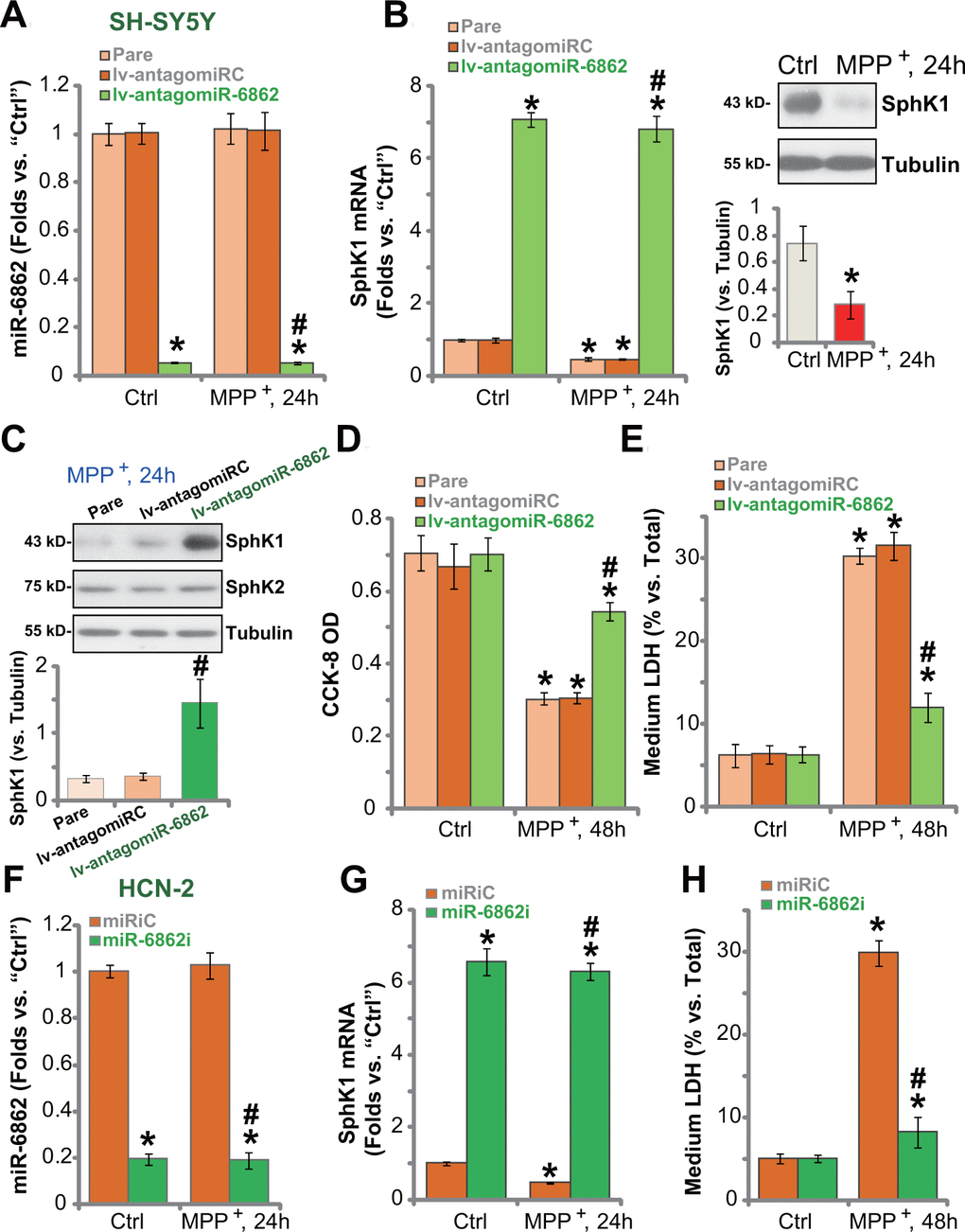
Figure 2. miR-6862 inhibition protects neuronal cells from MPP+. Parental control SH-SY5Y cells (“Pare”) as well as stable SH-SY5Y cells, expressing the lentiviral construct encoding the anti-sense of premiR-6862 (lv-antagomiR-6862) or the anti-sense control sequence (lv-antagomiRC), were treated with or without MPP+ (3 mM); Cells were then cultured for applied time periods, expression of miR-6862 (A) and SphK1 mRNA (B) was tested by qPCR assays, with SphK1 protein expression tested by Western blotting analyses (B, C); Cell viability and death were tested by the CCK-8 assay (D) and the LDH release assay (E), respectively. HCN-2 neuronal cells were transfected with 500 nM of miR-6862 inhibitor (miR-6862i) or the miR inhibitor control (miRiC) for 48h. Cells were then treated with or without MPP+ (3 mM) and cultured for indicated time periods, expression of miR-6862 (F) and SphK1 mRNA (G) was tested by qPCR, with cell death examined by medium LDH release assay (H). Bars stand for mean ± standard deviation (SD, n=5). * P < 0.05 vs. “Ctrl” treatment in “Pare” cells or “miRiC” cells. #P < 0.05 vs. MPP+ treatment in “Pare” cells or “miRiC” cells. Experiments in this figure were repeated five times, with the similar results obtained.
In line with our previous study [10], MPP+ treatment (3 mM, 48h) resulted in robust viability (CCK-8 OD) reduction (Figure 2D) and cell death (increased medium LDH release, Figure 2E) in parental SH-SY5Y cells. MPP+-induced cytotoxicity was however largely mitigated in lv-antagomiR-6862SH-SY5Y cells (Figure 2D, 2E). The lv-antagomiRC did not affect MPP+-induced cytotoxicity (Figure 2D, 2E).
In HCN-2 neuronal cells, transfection of miR-6862 inhibitor (miR-6862i) downregulated miR-6862 (Figure 2F) and induced SphK1 mRNA upregulation (Figure 2G) regardless of MPP+ treatment (Figure 2F, 2G). MPP+-induced HCN-2 cell death, evidenced by increased medium LDH release (Figure 2H), was largely inhibited by miR-6862i. Thus miR-6862 inhibition increased SphK1 expression and protected neuronal cells from MPP+-induced cytotoxicity.
miR-6862 inhibition attenuates MPP+-induced apoptosis in neuronal cells
Studies have shown that MPP+ inhibited SphK1 activity to provoke apoptosis activation in neuronal cells [20, 21]. We therefore tested whether miR-6862 inhibition could affect MPP+-induced neuronal cell apoptosis. In parental control SH-SY5Y cells, treatment with MPP+ significantly increased the activities of caspase-3 (Figure 3A) and caspase-9 (Figure 3B). It also caused single strand DNA (ssDNA) accumulation that indicates DNA breaks (Figure 3C). Mitochondrial depolarization, or JC-1 green monomers accumulation (increased JC-1 intensity at 488 nm), was detected in MPP+-treated SH-SY5Y cells (Figure 3D). Importantly, miR-6862 silencing by lv-antagomiR-6862 inhibited MPP+-induced caspase-3/-9 activation (Figure 3A, 3B), ssDNA accumulation (Figure 3C), and mitochondrial depolarization (Figure 3D) in SH-SY5Y cells. TUNEL staining assay results in Figure 3E showed that MPP+ induced significant apoptosis activation in SH-SY5Y cells. As TUNEL-positive nuclei ratio was significantly elevated in MPP+-treated cells (Figure 3E). Importantly, apoptosis activation by MPP+ was largely inhibited by lv-antagomiR-6862 (Figure 3E).
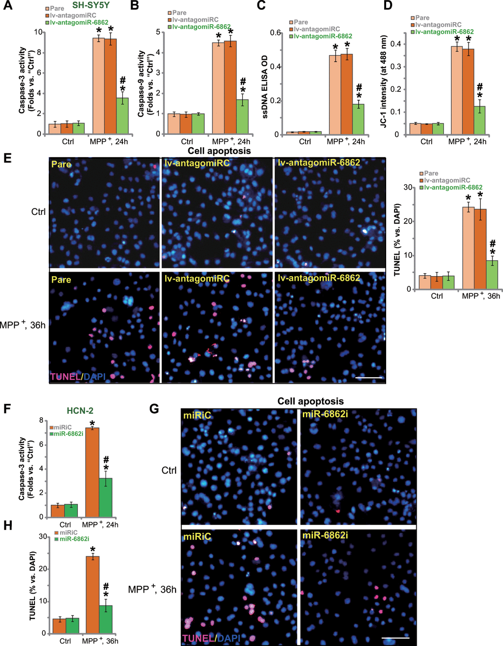
Figure 3. miR-6862 inhibition attenuates MPP+-induced apoptosis in neuronal cells. Parental control SH-SY5Y cells (“Pare”) as well as stable SH-SY5Y cells, expressing the lentiviral construct encoding the anti-sense of premiR-6862 (lv-antagomiR-6862) or the anti-sense control sequence (lv-antagomiRC), were treated with or without MPP+ (3 mM); Cells were then cultured for applied time periods, the caspase-3/-9 activity (A, B), single strand DNA (ssDNA) contents (C) and mitochondrial depolarization (recording JC-1 intensity at 488 nm, D) were tested. Cell apoptosis was tested by nuclear TUNEL staining assay (E). HCN-2 neuronal cells were transfected with 500 nM of miR-6862 inhibitor (miR-6862i) or the miR inhibitor control (miRiC) for 48h. Cells were then treated with or without MPP+ (3 mM) and cultured for indicated time periods, caspase-3 activity (F) and cell apoptosis (G, H) were tested similarly. Bars stand for mean ± standard deviation (SD, n=5). * P < 0.05 vs. “Ctrl” treatment in “Pare” cells or “miRiC” cells. #P < 0.05 vs. MPP+ treatment in “Pare” cells or “miRiC” cells. Experiments in this figure were repeated five times, with the similar results obtained. Scale bar= 100 μm (E, G).
In HCN-2 neuronal cells, transfection of the miR-6862 inhibitor (miR-6862i) largely inhibited MPP+-induced caspase-3 activation (Figure 3F) and apoptosis induction (TUNEL staining assay, Figure 3G, 3H). Collectively, miR-6862 inhibition alleviated MPP+-induced apoptosis in neuronal cells.
miR-6862 overexpression augments MPP+-induced neuronal cell death
Next, we studied whether miR-6862 overexpression would exert opposite functions and increase MPP+-induced neuronal cell death. In SH-SY5Y cells, forced miR-6862 overexpression by lv-premiR-6862 (see Figure 1) aggravated MPP+-induced SphK1 mRNA and protein downregulation (Figure 4A). MPP+-induced viability (CCK-8 OD) reduction (Figure 4B) and cell death (by recording medium LDH release, Figure 4C) were intensified in lv-premiR-6862 SH-SY5Y cells. Furthermore, with miR-6862 overexpression, MPP+-induced caspase-9 activation (Figure 4D), DNA breaks (ssDNA accumulation, Figure 4E) and mitochondrial depolarization (JC-1 green monomer intensity increase, Figure 4F) were augmented. Forced miR-6862 overexpression enhanced MPP+-induced apoptosis activation in SH-SY5Y cells (Figure 4G), which was reflected by increased TUNEL-positive nuclei ratio (Figure 4G).
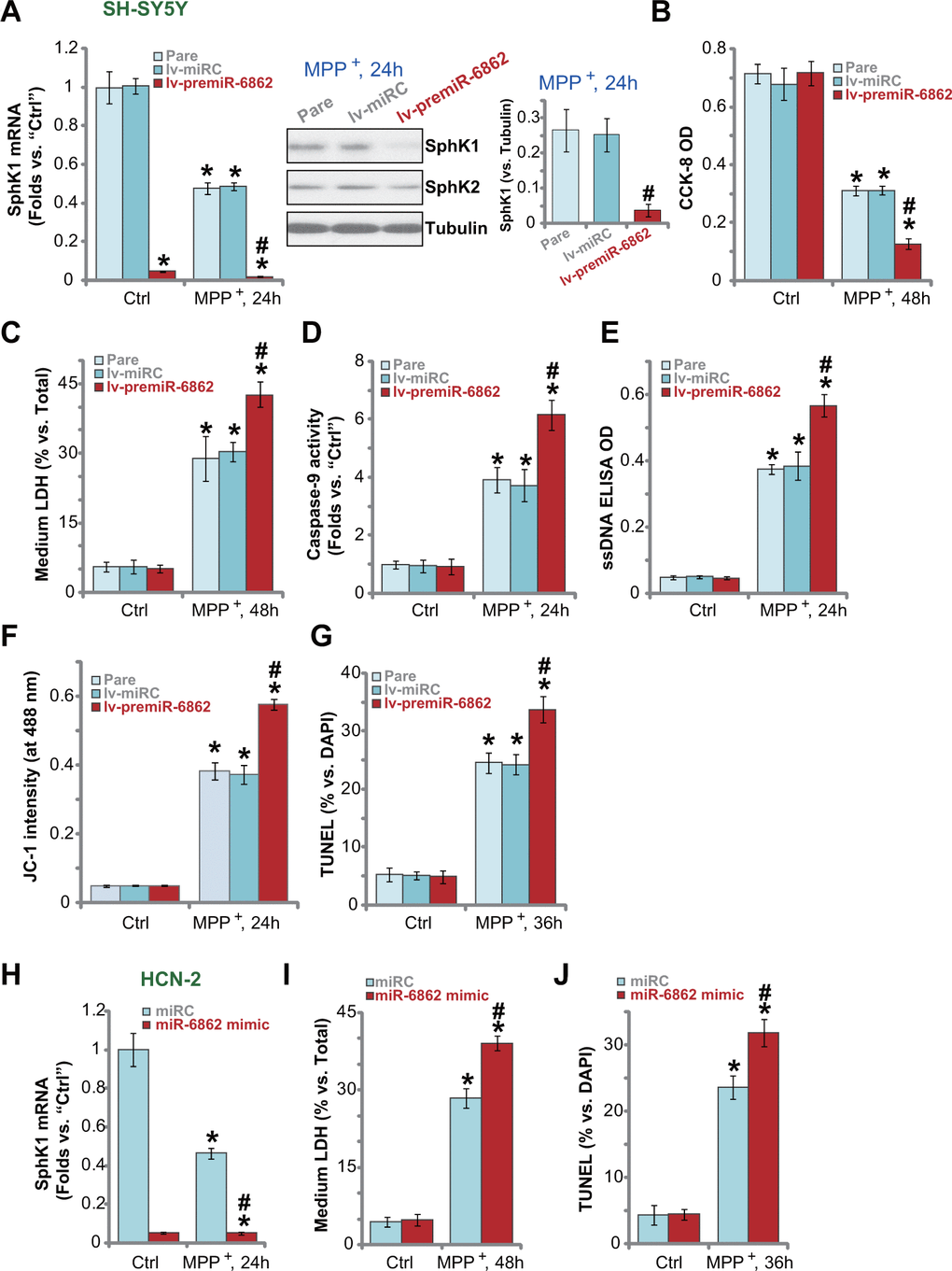
Figure 4. miR-6862 overexpression augments MPP+-induced neuronal cell death. Parental control SH-SY5Y cells (“Pare”) as well as stable SH-SY5Y cells, with the lentiviral construct encoding the premiR-6862 sequence (lv-premiR-6862) or the nonsense miRNA sequence (lv-miRC), were treated with or without MPP+ (3 mM), cells were then cultured for applied time periods, SphK1 mRNA and protein expression was tested (A); Cell viability and death were tested by CCK-8 assay (D) and LDH release assay (E), respectively; Caspase-9 activation (D), single strand DNA (ssDNA) contents (E) and mitochondrial depolarization (JC-1 dye assay, F) were tested, with cell apoptosis tested by nuclear TUNEL staining assay (G). HCN-2 neuronal cells were transfected with 500 nM of miR-6862 mimic or the miR control mimic (“miRC”) for 48h. Cells were then treated with or without MPP+ (3 mM) and cultured for indicated time periods, SphK1 mRNA expression (H), medium LDH contents (I), and cell apoptosis (by recording TUNEL-positive nuclei, (J) were tested. Bars stand for mean ± standard deviation (SD, n=5). * P < 0.05 vs. “Ctrl” treatment in “Pare” cells or “miRC” cells. #P < 0.05 vs. MPP+ treatment in “Pare” cells or “miRC” cells. Experiments in this figure were repeated five times, with the similar results obtained.
Transfection of miR-6862 mimic (500 nM) caused substantial SphK1 mRNA downregulation in HCN-2 neuronal cells with MPP+ treatment (Figure 4H). Consequently, MPP+-induced cell death (medium LDH release, Figure 4I) and apoptosis (reflected by TUNEL-positive nuclei ratio, Figure 4J) were augmented. Together, miR-6862 overexpression augmented MPP+-induced neuronal cell apoptosis.
SphK1 knockout intensifies MPP+-induced neuronal cell death
We further hypothesized that SphK1 knockout should mimic miR-6862 overexpression-induced actions. Therefore, a lentiCRISPR-GFP-SphK1-KO construct (from Dr. Yao at Nanjing Medical University [25]) was transduced to SH-SY5Y cells. Stable cells were established (koSphK1 cells, see Methods). SphK1 mRNA (Figure 5A) and protein (Figure 5B) expression were depleted in koSphK1 cells. As compared to control cells with empty vector (“Cas9-C”), MPP+-induced viability reduction was intensified in koSphK1 SH-SY5Y cells (Figure 5C). Furthermore, in koSphK1 cells, MPP+-induced cell death (medium LDH release, Figure 5D) and apoptosis (by recording TUNEL-positive nuclei ratio, Figure 5E) were aggravated. Therefore, CRISPR/Cas9-induced SphK1 KO intensified MPP+-induced neuronal cell death.
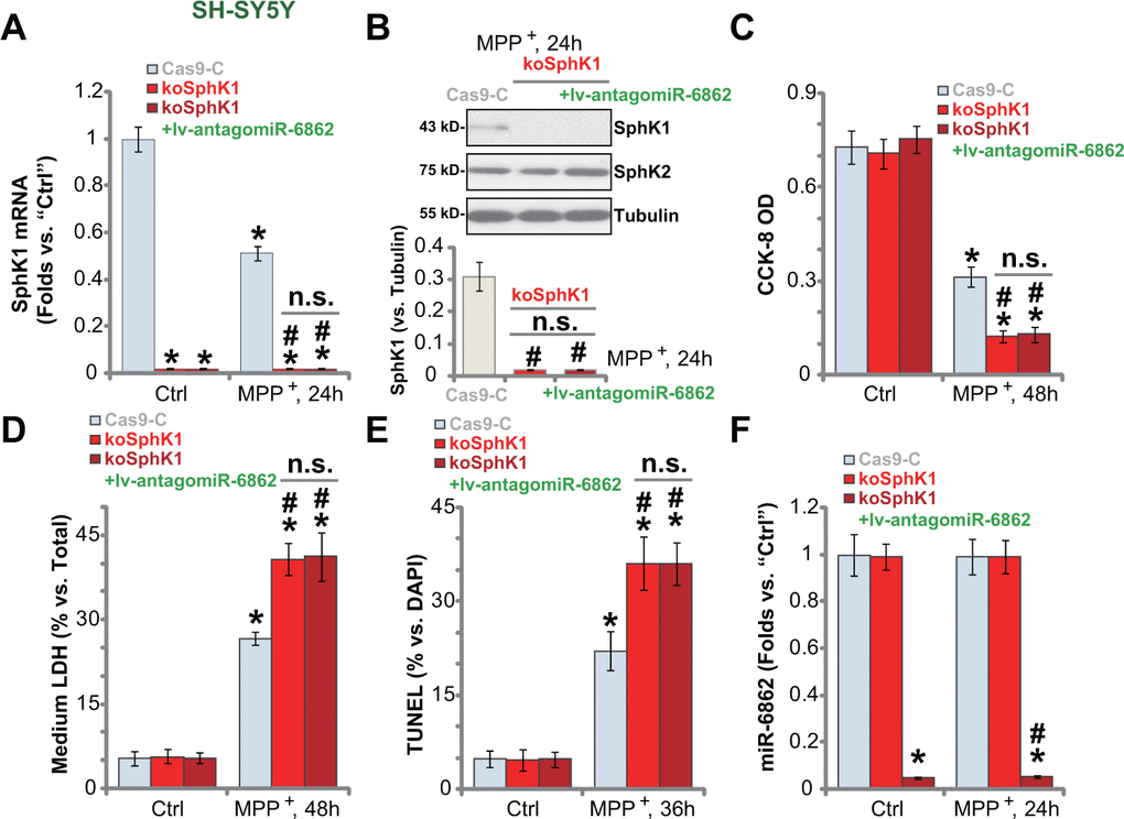
Figure 5. SphK1 knockout intensifies MPP+-induced neuronal cell death. Stable SH-SY5Y cells expressing the lentiCRISPR-GFP-SphK1-KO construct (“koSphK1” cells) were further transduced with or without the lentiviral construct encoding the anti-sense of premiR-6862 (lv-antagomiR-686), control cells were transduced with lentiCRISPR-GFP empty vector (“Cas9-C”); Cells were treated with or without MPP+ (3 mM) and then cultured for applied time periods, SphK1 mRNA and protein expression was tested (A, B); Cell viability and death were tested by CCK-8 (C) and LDH release (D) assays, respectively; Cell apoptosis was examined by nuclear TUNEL staining assay (E). miR-6862 expression was shown (F). Bars stand for mean ± standard deviation (SD, n=5). * P < 0.05 vs. “Ctrl” treatment in “Cas9-C” cells. #P < 0.05 vs. MPP+ treatment in “Cas9-C” cells. “n.s.” stands for non-statistical difference. Experiments in this figure were repeated five times, with the similar results obtained.
Importantly, though lv-antagomiR-6862 silenced miR-6862 (Figure 5F) in koSphK1 cells, it failed to offer neuroprotective activity against MPP+. Specifically, MPP+-induced viability reduction (Figure 5C), cell death (Figure 5D) and apoptosis (Figure 5E) were not alleviated by lv-antagomiR-6862 in koSphK1 SH-SY5Y cells. These results suggested that miR-6862 inhibition protects neuronal cells from MPP+-induced apoptosis through upregulating SphK1.
Discussion
The loss of DA neurons in the midbrain is a key pathological characteristics of PD progression, and it could be due to mitochondrial dysfunction, free radicals (ROS) accumulation, sustained oxidative injury, as well as abnormal protein aggregation and inflammatory lesions [5, 29]. Of particular relevance, mitochondrial dysfunction, causing ROS accumulation and oxidative injury, is one primary cause of DA neuronal cell death [5, 29].
The postmortem brain analyses and animal model studies have revealed that miRNA dysregulation is associated with PD [30, 31] and other neurodegenerative disorders such as Alzheimer's disease and Huntington's disease [32, 33]. Understanding miRNAs and their targeted genes in neurodegenerative diseases should help to identify novel genes for neuronal functions and to reveal the underlying molecular mechanisms underlying these diseases [32, 33]. Since miRNAs regulating genes and pathways that are associated with PD are altered in PD, it is useful in the diagnose of PD and even different PD subtypes [30, 31]. Furthermore, miRNAs are important in regulating neuronal cell death by MPP+.
Datta et al., have shown that microRNA-7 (miR-7) prevented MPP+-induced cell death in SH-SY5Y cells and differentiated human neural progenitor ReNcell VM cells [34]. In addition, miR-7 downregulated RelA, a member of the nuclear factor κB (NF-κB) family of transcription factors, and augmented neuronal glucose transporter 3 (Glut3) to promote glycolysis in SH-SY5Y cells and ReNcell VM cells [34]. Mao et al., found that miR-21 downregulation was able to attenuate MPP+-induced MES23.5 cell death by apoptosis inhibition and reactive oxygen species (ROS) scavenging [35]. In SH-SY5Y cells and primary dopaminergic neurons, astrocyte-derived exosomal miR-200a-3p downregulated MKK4 (mitogen-activated protein kinase kinase 4) and prevented MPP+-induced apoptotic cell death [36]. miR-6862 is a relatively novel miRNA with its functions largely unknown. Fuhrmannet al., demonstrated that miR-6862 silenced nuclear receptor coactivator 4 (NCOA4) to inhibit mitochondrial ferritin (FTMT) formation [37]. Zhu et al., reported that miR-6862 expression is dysregulated in lung adenocarcinoma. Here we identified miR-6862 as a novel SphK1-targeting miRNA in neuronal cells. MiR-6862 locates in the cytoplasm of SH-SY5Y neuronal cells and directly binds to SphK1 mRNA. Ectopic overexpression of miR-6862 decreased SphK1 3’-UTR luciferase reporter activity and downregulated its expression in neuronal cells. miR-6862 inhibition, by lv-antagomiR-6862 construct or miR-6862 inhibitor, exerted opposite activity and elevated SphK1 expression in neuronal cells.
SphK1 converts sphingosine to S1P, the bioactive lipids with important functions in cell survival, differentiation, migration, and trafficking [14, 38]. SphK1 downregulation was detected in MPP+-treated SH-SY5Y neuronal cells [21]. Exogenous S1P addition would activate SphK1 and inhibited MPP+-induced neuronal cell apoptosis [20, 21]. K6PC-5, a SphK1 activator, protected SH-SY5Y neuronal cells from oxygen glucose deprivation/re-oxygenation-induced oxidative stress [22]. Here in SH-SY5Y and HCN-2 neuronal cells, MPP+-induced cell death was attenuated by miR-6862 inhibition with either lv-antagomiR-6862 or miR-6862 inhibitor. Conversely, ectopic overexpression of miR-6862 further downregulated SphK1 and augmented MPP+-induced neuronal cell apoptosis. Thus, SphK1 elevation by miR-6862 inhibition protected neuronal cells from MPP+-induced apoptosis.
Importantly, CRISPR/Cas9-induced SphK1 KO augmented MPP+-induced neuronal cell death, which mimicked miR-6862 overexpression-induced activity. Significantly, in koSphK1 SH-SY5Y neuronal cells, exogenously miR-6862 inhibition by lv-antagomiR-6862 failed to alleviate MPP+-induced apoptosis. These results indicated that SphK1 should be the primary target of miR-6862 in neuronal cells. Moreover, miR-6862 inhibition-induced neuronal cell protection against MPP+ should be through elevating SphK1 expression.
However, the findings of antagomiR-6862-induced neuronal cell protection in vitro cannot be simply translated in vivo. The in vivo experiments are regarded as the gold standard for PD studies [39]. The potential effect of antagomiR-6862 remains to be evaluated in MPTP-induced PD animal model and possibly in genetic-based PD animal models. Also, only established neuronal cell lines, SH-SY5Y and HCN-2, were tested in this study. Our findings have to be further verified in primary DA neurons. In addition, miR-6862 expression was unchanged in MPP+-treated cells. Further exploring the expression of miR-6862 in PD postmortem brain tissues and PD animal models would be necessary.
In summary, we identified miR-6862 as a novel SphK1-targeting miRNA. miR-6862 inhibition upregulated SphK1 and protected neuronal cells from MPP+-induced damage. Therefore, SphK1 elevation induced bymiR-6862 inhibition could be a novel genetic strategy to protect DA neurons against oxidative injury.
Materials and Methods
Reagents and antibodies
MPP+, puromycin, polybrene and CCK-8 assay kit were obtained from Sigma-Aldrich (St. Louis, Mo). The antibodies were all provided by Cell Signaling Tech (Danvers, MA) and Abcam (Shanghai, China). Lipofectamine 2000 and other transfection reagents were provided by Thermo-Fisher Invitrogen (Shanghai, China). All the primers, sequences, and viral constructs were provided by Shanghai Genechem Co. (Shanghai, China) unless otherwise mentioned.
Cell culture
Culturing the differentiated SH-SY5Y neuronal cells was reported before [10, 40]. The HCN-2 neuronal cell line was purchased from the Cell Bank of Shanghai Institute of Biological Science (Shanghai, China). HCN-2 cells were cultured in DMEM with 10% FBS.MPP+ was dissolved in PBS to obtain 30 mM MPP+ stock solution, and was then filtrated and stored in dark at -20° C. Neuronal cells with applied genetic modifications were treated with MPP+ (3 mM). This concentration was chosen based on our previous studies [10, 41].
miR-6862 overexpression or inhibition
GV369 lentiviral constructs encoding the miR-6862 precursor sequence (premiR-6862, sequence listed in Table 2) or the anti-sense sequence (anti-premiR-6862) were synthesized by Genechem (Shanghai, China). Each was transfected in HEK-293T cells along with the lentivirus package plasmids (Genechem) to generate the premiR-6862 expression lentivirus (“lv-premiR-6862”) and the premiR-6862 anti-sense lentivirus (“lv-antagomiR-6862”). Neuronal cells were cultured into six well-tissue plates (at 2 × 10⁵ cells per well)under the polybrene-containing medium, and lentivirus was then added. Puromycin (5.0 μg/mL) was further added to select stable cells, and mature miR-6862 expression (sequence listed in Table 2) was examined by qPCR.
Transfection of miR mimic
SH-SY5Y and HCN-2 neuronal cells were cultured into six well-tissue plates (at 2 × 10⁵ cells per well) and transfected with 500 nM of the applied miR mimic or miR inhibitor through a Lipofectamine 2000 protocol [42].
SphK1 3'-UTR activity assay
Briefly, SphK1 3’-UTR containing the putative binding sites of miR-6862(at position 113-120) was provided by Dr. Yao [25]. It was inserted into a firefly luciferase reporter vector pGL4.13 (luc2/SV40) [25]. SH-SY5Y neuronal cells were cultured into six well-tissue plates (at 2 × 10⁵ cells per well) and transfected with the plasmid as well as the Renilla luciferase reporter vector and pRL-SV40 [25] (by Lipofectamine 2000). Cells with the applied genetic modifications were subjected to SphK1 3'-UTR luciferase activity assay using a Promega kit [42].
RNA-Pull down assay
RNA-Pull down was analyzed by a Pierce Magnetic RNA Pull-Down Kit [43, 44]. Briefly, SH-SY5Y neuronal cells were cultured into six well-tissue plates (at 2 × 10⁵ cells per well) and transfected with biotinylated miR-6862 mimic (100 nmol/L) for 48h [44]. The streptavidin-coated magnetic beads were added to total cell lysates to pull-down biotin-captured miR-6862-bound RNA complex [43]. SphK1 mRNA expression was tested by qPCR with level normalized (% of input controls).
Cell viability
Neuronal cells were plated into 96 well-tissue plates (at 3, 000 cells per well). Following the applied MPP+ treatment, a CCK-8 assay kit was utilized to test cell viability with the attached protocol. CCK-8’s optical density (OD) was tested at the test-wavelength of 550 nm.
Quantitative real-time reverse transcriptase polymerase chain reaction (qPCR)
The detailed protocols of qPCR were described in our previous study [10]. Briefly, TRIzol reagents (Sigma) were utilized to extract total cellular RNA, which was then reversely transcribed to cDNA. A TOYOBO ReverTra Ace qPCR kit (Tokyo, Japan) was applied for qPCR under the ABI Prism 7500H fast Real-Time PCR system (Foster City, CA). The methods for mRNA data quantification and normalization to GAPDH were described before [10]. A TransStartTM SYBR Green qPCR Supermix (TransGen Biotech) was utilized to examine miR-6862 expression, and the results were normalized to U6 RNA. The mRNA primers were listed in Table 2.
Lactate dehydrogenase (LDH) assay
Neuronal cells were plated in six well-tissue plates. Following the applied MPP+ treatment, LDH contents were tested by a simple two-step LDH detection kit (Promega, Shanghai, China). LDH contents in the medium were always normalized to total LDH contents.
Western blotting
Neuronal cells were plated into six well-tissue plates (at 2 × 10⁵ cells per well). Following the applied treatment, cell lysates were obtained. Detailed protocols of Western blotting were described before [10]. Data quantification was performed through ImageJ software (NIH).
Caspase activity
Neuronal cells were plated into six well-tissue plates (at 2 × 10⁵ cells per well). Following the MPP+ treatment, cell lysates were obtained. The relative activities of caspase-3 and caspase-9 were tested by fluorometric caspase assay kits (Beyotime Biotechnology, Wuxi, China) [45] with 20 μg lysates per sample. The caspase-3 or the caspase-9 p-nitroaniline (pNA) absorbance was detected at the wavelength of 405 nm.
Single strand DNA (ssDNA) ELISA assay. Neuronal cells were seeded into six-well plates. Following the applied MPP+ treatment, ssDNA contents were tested using a ApoStrandTM ELISA kit (BIOMOL International, Plymouth Meeting, PA).
TUNEL staining
Neuronal cells were plated into six well-tissue plates (at 2 × 10⁵ cells per well). Following MPP+ treatment, cell apoptosis was measured via a TUNEL [terminal deoxynucleotidyl transferase (TdT)-mediated dUTP nick end labeling] In Situ Cell Death Detection Kit (Roche, Shanghai, China). TUNEL-positive nuclei percentage (% vs. DAPI), from 500 cells per treatment in five random views (1: 100 magnification), was always calculated.
Mitochondrial depolarization assay
Neuronal cells were plated into 96 well-tissue plates (at 3, 000 cells per well). Following the applied MPP+ treatment, cells were stained with the mito-dye JC-1. JC-1 green fluorescence intensity (at 488 nm) was recorded. The representative JC-1 images were presented as well.
SphK1 knockout (KO)
A lentiCRISPR-GFP construct encoding the small guide RNA (sgRNA) against human SphK1 was provided by Dr. Yao at Nanjing Medical University [25]. SH-SY5Y neuronal cells were cultured into six well-tissue plates and transfected with SphK1-KO construct. FACS-mediated GFP sorting were utilized to select transfected cells. Cells were then distributed into 96-well plates. Single stable cells with SphK1 KO were further screened by qPCR. Control SH-SY5Y cells were transfected with the empty vector [25].
Statistics
Data were presented as mean ±standard deviation (SD). Statistical differences were analyzed by one-way analysis of variance (ANOVA) followed by multiple comparisons performed with post hoc Bonferroni test (SPSS, 23.0, SPSS Co. Chicago, CA). A two-tailed unpaired T test was utilized (Excel 2007) when comparing the difference between two treatment groups. Values with P < 0.05 were considered statistically significant.
Author Contributions
GX performed all neuronal culture and signaling studies. YL helped to established and verified constructs virus and sequences. JC performed all cell death studies. ZZ, JZ and WD: Study conception and design, and data analysis, Figure organization, involved in drafting the article and revising it critically for important intellectual content, and with final approval of the version submitted to the journal.
Conflicts of Interest
None of the authors have any conflicts of interest.
Funding
This work was supported by funds from a special project “diagnostic and therapeutic technology of key clinical diseases” of Suzhou (LCZX201404) and a grant from NSFC of China (81974388).
References
- 1. Ascherio A, Schwarzschild MA. The epidemiology of Parkinson’s disease: risk factors and prevention. Lancet Neurol. 2016; 15:1257–72. https://doi.org/10.1016/S1474-4422(16)30230-7 [PubMed]
- 2. de Lau LM, Breteler MM. Epidemiology of Parkinson’s disease. Lancet Neurol. 2006; 5:525–35. https://doi.org/10.1016/S1474-4422(06)70471-9 [PubMed]
- 3. Van Den Eeden SK, Tanner CM, Bernstein AL, Fross RD, Leimpeter A, Bloch DA, Nelson LM. Incidence of Parkinson’s disease: variation by age, gender, and race/ethnicity. Am J Epidemiol. 2003; 157:1015–22. https://doi.org/10.1093/aje/kwg068 [PubMed]
- 4. McNaught KS, Olanow CW, Halliwell B, Isacson O, Jenner P. Failure of the ubiquitin-proteasome system in Parkinson’s disease. Nat Rev Neurosci. 2001; 2:589–94. https://doi.org/10.1038/35086067 [PubMed]
- 5. Lotharius J, Brundin P. Pathogenesis of Parkinson’s disease: dopamine, vesicles and alpha-synuclein. Nat Rev Neurosci. 2002; 3:932–42. https://doi.org/10.1038/nrn983 [PubMed]
- 6. Irwin DJ, Lee VM, Trojanowski JQ. Parkinson’s disease dementia: convergence of α-synuclein, tau and amyloid-β pathologies. Nat Rev Neurosci. 2013; 14:626–36. https://doi.org/10.1038/nrn3549 [PubMed]
- 7. Al Shahrani M, Heales S, Hargreaves I, Orford M. Oxidative stress: mechanistic insights into inherited mitochondrial disorders and Parkinson’s disease. J Clin Med. 2017; 6:100. https://doi.org/10.3390/jcm6110100 [PubMed]
- 8. Gaki GS, Papavassiliou AG. Oxidative stress-induced signaling pathways implicated in the pathogenesis of Parkinson’s disease. Neuromolecular Med. 2014; 16:217–30. https://doi.org/10.1007/s12017-014-8294-x [PubMed]
- 9. Perier C, Bové J, Vila M. Mitochondria and programmed cell death in Parkinson’s disease: apoptosis and beyond. Antioxid Redox Signal. 2012; 16:883–95. https://doi.org/10.1089/ars.2011.4074 [PubMed]
- 10. Zhu JL, Wu YY, Wu D, Luo WF, Zhang ZQ, Liu CF. SC79, a novel Akt activator, protects dopaminergic neuronal cells from MPP+ and rotenone. Mol Cell Biochem. 2019; 461:81–89. https://doi.org/10.1007/s11010-019-03592-x [PubMed]
- 11. Ito K, Eguchi Y, Imagawa Y, Akai S, Mochizuki H, Tsujimoto Y. MPP+ induces necrostatin-1- and ferrostatin-1-sensitive necrotic death of neuronal SH-SY5Y cells. Cell Death Discov. 2017; 3:17013. https://doi.org/10.1038/cddiscovery.2017.13 [PubMed]
- 12. Kong XC, Zhang D, Qian C, Liu GT, Bao XQ. FLZ, a novel HSP27 and HSP70 inducer, protects SH-SY5Y cells from apoptosis caused by MPP(+). Brain Res. 2011; 1383:99–107. https://doi.org/10.1016/j.brainres.2011.01.093 [PubMed]
- 13. Choi WS, Yoon SY, Oh TH, Choi EJ, O’Malley KL, Oh YJ. Two distinct mechanisms are involved in 6-hydroxydopamine- and MPP+-induced dopaminergic neuronal cell death: role of caspases, ROS, and JNK. J Neurosci Res. 1999; 57:86–94. https://doi.org/10.1002/(SICI)1097-4547(19990701)57:1<86::AID-JNR9>3.0.CO;2-E [PubMed]
- 14. Maceyka M, Harikumar KB, Milstien S, Spiegel S. Sphingosine-1-phosphate signaling and its role in disease. Trends Cell Biol. 2012; 22:50–60. https://doi.org/10.1016/j.tcb.2011.09.003 [PubMed]
- 15. Edmonds Y, Milstien S, Spiegel S. Development of small-molecule inhibitors of sphingosine-1-phosphate signaling. Pharmacol Ther. 2011; 132:352–60. https://doi.org/10.1016/j.pharmthera.2011.08.004 [PubMed]
- 16. Shida D, Takabe K, Kapitonov D, Milstien S, Spiegel S. Targeting SphK1 as a new strategy against cancer. Curr Drug Targets. 2008; 9:662–73. https://doi.org/10.2174/138945008785132402 [PubMed]
- 17. Okada T, Kajimoto T, Jahangeer S, Nakamura S. Sphingosine kinase/sphingosine 1-phosphate signalling in central nervous system. Cell Signal. 2009; 21:7–13. https://doi.org/10.1016/j.cellsig.2008.07.011 [PubMed]
- 18. Lee JY, Han SH, Park MH, Baek B, Song IS, Choi MK, Takuwa Y, Ryu H, Kim SH, He X, Schuchman EH, Bae JS, Jin HK. Neuronal SphK1 acetylates COX2 and contributes to pathogenesis in a model of Alzheimer’s disease. Nat Commun. 2018; 9:1479. https://doi.org/10.1038/s41467-018-03674-2 [PubMed]
- 19. Meng H, Yuan Y, Lee VM. Loss of sphingosine kinase 1/S1P signaling impairs cell growth and survival of neurons and progenitor cells in the developing sensory ganglia. PLoS One. 2011; 6:e27150. https://doi.org/10.1371/journal.pone.0027150 [PubMed]
- 20. Pyszko JA, Strosznajder JB. The key role of sphingosine kinases in the molecular mechanism of neuronal cell survival and death in an experimental model of Parkinson’s disease. Folia Neuropathol. 2014; 52:260–69. https://doi.org/10.5114/fn.2014.45567 [PubMed]
- 21. Pyszko J, Strosznajder JB. Sphingosine kinase 1 and sphingosine-1-phosphate in oxidative stress evoked by 1-methyl-4-phenylpyridinium (MPP+) in human dopaminergic neuronal cells. Mol Neurobiol. 2014; 50:38–48. https://doi.org/10.1007/s12035-013-8622-4 [PubMed]
- 22. Liu H, Zhang Z, Xu M, Xu R, Wang Z, Di G. K6PC-5 activates SphK1-Nrf2 signaling to protect neuronal cells from oxygen glucose deprivation/re-oxygenation. Cell Physiol Biochem. 2018; 51:1908–20. https://doi.org/10.1159/000495716 [PubMed]
- 23. Treiber T, Treiber N, Meister G. Regulation of microRNA biogenesis and its crosstalk with other cellular pathways. Nat Rev Mol Cell Biol. 2019; 20:5–20. https://doi.org/10.1038/s41580-018-0059-1 [PubMed]
- 24. Nie JH, Li TX, Zhang XQ, Liu J. Roles of non-coding RNAs in normal human brain development, brain tumor, and neuropsychiatric disorders. Noncoding RNA. 2019; 5:36. https://doi.org/10.3390/ncrna5020036 [PubMed]
- 25. Yao C, Ruan JW, Zhu YR, Liu F, Wu HM, Zhang Y, Jiang Q. The therapeutic value of the SphK1-targeting microRNA-3677 in human osteosarcoma cells. Aging (Albany NY). 2020; 12:5399–410. https://doi.org/10.18632/aging.102961 [PubMed]
- 26. Zhou Y, Han Y, Zhang Z, Shi Z, Zhou L, Liu X, Jia X. MicroRNA-124 upregulation inhibits proliferation and invasion of osteosarcoma cells by targeting sphingosine kinase 1. Hum Cell. 2017; 30:30–40. https://doi.org/10.1007/s13577-016-0148-4 [PubMed]
- 27. Zhang H, Wang Q, Zhao Q, Di W. MiR-124 inhibits the migration and invasion of ovarian cancer cells by targeting SphK1. J Ovarian Res. 2013; 6:84. https://doi.org/10.1186/1757-2215-6-84 [PubMed]
- 28. Agarwal V, Bell GW, Nam JW, Bartel DP. Predicting effective microRNA target sites in mammalian mRNAs. Elife. 2015; 4:e05005. https://doi.org/10.7554/eLife.05005 [PubMed]
- 29. Abou-Sleiman PM, Muqit MM, Wood NW. Expanding insights of mitochondrial dysfunction in Parkinson’s disease. Nat Rev Neurosci. 2006; 7:207–19. https://doi.org/10.1038/nrn1868 [PubMed]
- 30. Fyfe I. MicroRNAs - diagnostic markers in Parkinson disease? Nat Rev Neurol. 2020; 16:65. https://doi.org/10.1038/s41582-019-0305-y [PubMed]
- 31. Goh SY, Chao YX, Dheen ST, Tan EK, Tay SS. Role of MicroRNAs in Parkinson’s disease. Int J Mol Sci. 2019; 20:5649. https://doi.org/10.3390/ijms20225649 [PubMed]
- 32. Chan AW, Kocerha J. The path to microRNA therapeutics in psychiatric and neurodegenerative disorders. Front Genet. 2012; 3:82. https://doi.org/10.3389/fgene.2012.00082 [PubMed]
- 33. Lau P, de Strooper B. Dysregulated microRNAs in neurodegenerative disorders. Semin Cell Dev Biol. 2010; 21:768–73. https://doi.org/10.1016/j.semcdb.2010.01.009 [PubMed]
- 34. Chaudhuri AD, Kabaria S, Choi DC, Mouradian MM, Junn E. MicroRNA-7 promotes glycolysis to protect against 1-methyl-4-phenylpyridinium-induced cell death. J Biol Chem. 2015; 290:12425–34. https://doi.org/10.1074/jbc.M114.625962 [PubMed]
- 35. Mao H, Ding L. Downregulation of miR-21 suppresses 1-methyl-4-phenylpyridinium-induced neuronal damage in MES23.5 cells. Exp Ther Med. 2019; 18:2467–74. https://doi.org/10.3892/etm.2019.7853 [PubMed]
- 36. Shakespear N, Ogura M, Yamaki J, Homma Y. Astrocyte-derived exosomal microRNA miR-200a-3p prevents MPP+-induced apoptotic cell death through down-regulation of MKK4. Neurochem Res. 2020; 45:1020–33. https://doi.org/10.1007/s11064-020-02977-5 [PubMed]
- 37. Fuhrmann DC, Mondorf A, Beifuß J, Jung M, Brüne B. Hypoxia inhibits ferritinophagy, increases mitochondrial ferritin, and protects from ferroptosis. Redox Biol. 2020; 36:101670. https://doi.org/10.1016/j.redox.2020.101670 [PubMed]
- 38. Alemany R, van Koppen CJ, Danneberg K, Ter Braak M, Meyer Zu Heringdorf D. Regulation and functional roles of sphingosine kinases. Naunyn Schmiedebergs Arch Pharmacol. 2007; 374:413–28. https://doi.org/10.1007/s00210-007-0132-3 [PubMed]
- 39. Blesa J, Phani S, Jackson-Lewis V, Przedborski S. Classic and new animal models of Parkinson’s disease. J Biomed Biotechnol. 2012; 2012:845618. https://doi.org/10.1155/2012/845618 [PubMed]
- 40. Knaryan VH, Samantaray S, Park S, Azuma M, Inoue J, Banik NL. SNJ-1945, a calpain inhibitor, protects SH-SY5Y cells against MPP(+) and rotenone. J Neurochem. 2014; 130:280–90. https://doi.org/10.1111/jnc.12629 [PubMed]
- 41. Zheng J, Zhu JL, Zhang Y, Zhang H, Yang Y, Tang DR, Sun J. PGK1 inhibitor CBR-470-1 protects neuronal cells from MPP+. Aging (Albany NY). 2020; 12:13388–99. https://doi.org/10.18632/aging.103443 [PubMed]
- 42. Liu YY, Chen MB, Cheng L, Zhang ZQ, Yu ZQ, Jiang Q, Chen G, Cao C. microRNA-200a downregulation in human glioma leads to Gαi1 over-expression, Akt activation, and cell proliferation. Oncogene. 2018; 37:2890–902. https://doi.org/10.1038/s41388-018-0184-5 [PubMed]
- 43. Wang R, Zhang S, Chen X, Li N, Li J, Jia R, Pan Y, Liang H. CircNT5E acts as a sponge of miR-422a to promote glioblastoma tumorigenesis. Cancer Res. 2018; 78:4812–25. https://doi.org/10.1158/0008-5472.CAN-18-0532 [PubMed]
- 44. Wang K, Long B, Liu F, Wang JX, Liu CY, Zhao B, Zhou LY, Sun T, Wang M, Yu T, Gong Y, Liu J, Dong YH, et al. A circular RNA protects the heart from pathological hypertrophy and heart failure by targeting miR-223. Eur Heart J. 2016; 37:2602–11. https://doi.org/10.1093/eurheartj/ehv713 [PubMed]
- 45. Li KR, Zhang ZQ, Yao J, Zhao YX, Duan J, Cao C, Jiang Q. Ginsenoside rg-1 protects retinal pigment epithelium (RPE) cells from cobalt chloride (CoCl2) and hypoxia assaults. PLoS One. 2013; 8:e84171. https://doi.org/10.1371/journal.pone.0084171 [PubMed]