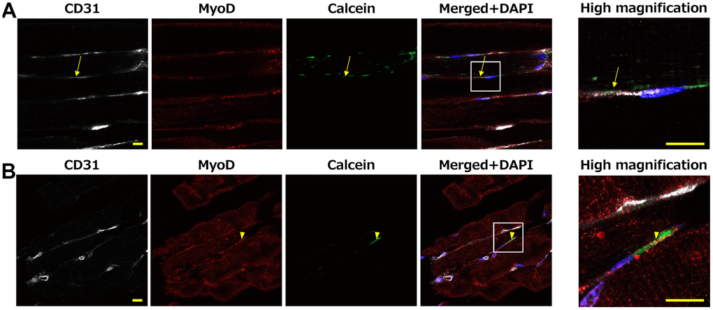
Figure 8.Mouse muscle at 10 minutes after Calcein loaded WBC intravenous injection. Calcein signals were observed in the muscle tissue, including CD31-positive endothelial cells (A: arrow) and MyoD-positive satellite cells (B: arrowhead). High-magnification images of the white squares are shown in right panel. Scale bar = 10 μm (A, B.; left and right panels).