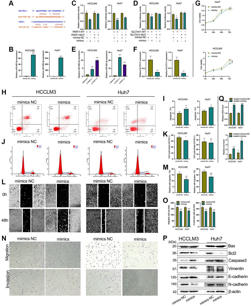
Figure 7.PART1 promotes HCC progression and inhibits ferroptosis by sponging miR-490-3p. (A) The interaction of miR-490-3p with PART1 and SLC7A11 predicted by ENCORI and LncBase v2 database. (B) The relative expression of miR-490-3p detected by RT-qPCR in HCCLM3 and Huh7 cells treated with miR-490-3p mimics or mimics NC. (C, D) Results of dual-luciferase reporter assay for miRNA-490-3p targeting PART1 and SLC7A11 mRNA 3'-UTR in HCCLM3 and Huh7 cells, respectively. (E) The relative expression of miR-490-3p in HCCLM3 and Huh7 cells transfected with siRNA-PART1.1 and -PART1.3. (F) The relative expression of SLC7A11 detected by qPCR in HCCLM3 and Huh7 cells treated with miR-490-3p mimics or mimics NC. (G) The cell proliferation was measured by the CCK-8 assays in HCCLM3 and Huh7 cells treated with miR-490-3p mimics or mimics NC. (H–K) The cell apoptosis and cell cycle were assessed by flow cytometry analysis in HCCLM3 and Huh7 cells treated with miR-490-3p mimics or mimics NC. (L, M) The migration was examined by wound healing assays in HCCLM3 and Huh7 cells treated with miR-490-3p mimics or mimics NC. (N, O) The cell migration and invasion were detected by Transwell assays (40 × objective lens) in HCCLM3 and Huh7 cells treated with miR-490-3p mimics or mimics NC. (P) The apoptosis and EMT-related proteins were detected by Western blotting. (Q) The relative levels of MDA and iron in HCCLM3 and Huh7 cells treated with miR-490-3p mimics or mimics NC in the erastin-induced ferroptosis assay. *p < 0.05, **p < 0.01, ***p < 0.001.