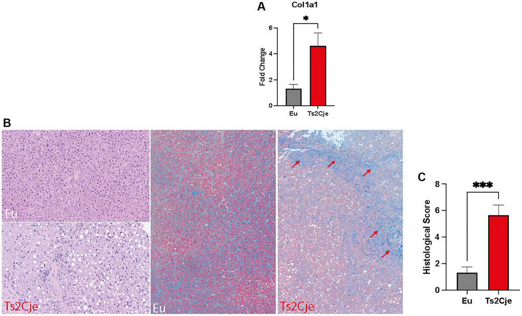
Figure 4.DS mice show a marked increase in fibrotic markers. (A) Col1a1 expression level is increased in Ts2Cje livers. Representative histogram of the real-time PCR against Col1a1 on the liver extract obtained from 12-months old Eu and Ts2Cje mice. GAPDH was used as housekeeping gene. (B) Ts2Cje liver section shows an increased fibrotic level. Representative histological images of the rat livers from control and Ts2Cje group. (Top left panel) Mouse liver from control group showing absence of steatosis (score 0) and mild periportal inflammation (score 1) (hematoxylin and eosin; original magnification 100x). (Bottom left panel) Mouse liver from Ts2Cje group exhibiting moderate steatosis (score 2) and moderate portal inflammation (score 2) (hematoxylin and eosin; original magnification 150x). (Mid panel) Mice liver from control group showing absence of fibrosis (score 0) (Masson’s trichrome; original magnification 50x). (Right panel) Mice liver from Ts2Cje group exhibiting diffuse fibrosis (score 3) with fibrous bridging (arrows) (Masson’s trichrome; original magnification 50x). (C) Histological score quantified as in B. Histograms are representative of four different experiments (*P ≤ 0.05; ***P ≤ 0.001).