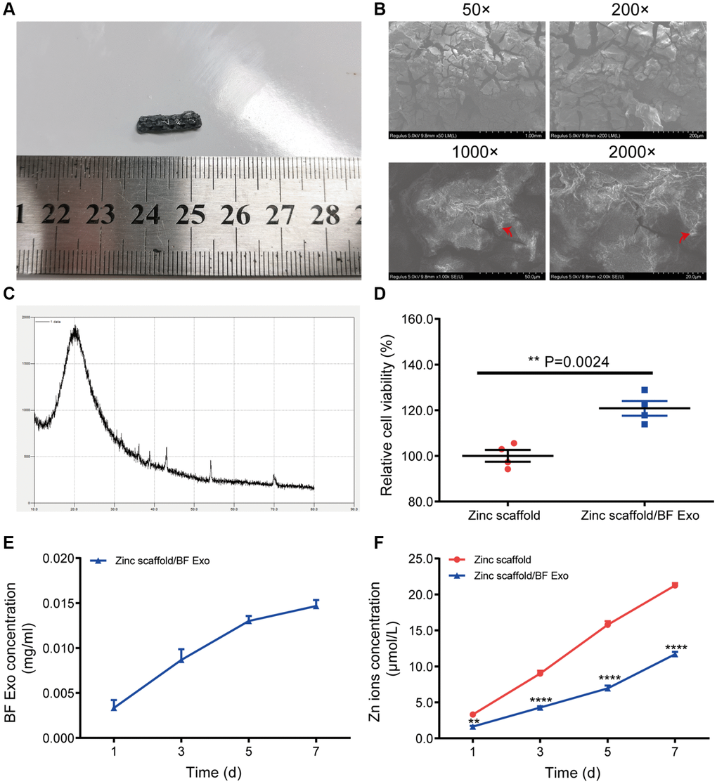
Figure 3.Preparation of zinc scaffold/BF Exo composite implant. (A) Solid images of degradable zinc stent/BF Exo composite implant; (B) Electron microscopy images of degradable zinc stent/BF Exo composite implant at 50, 200, 1000, and 2000 times magnification, with red arrows indicating small amounts of exosomes encapsulated in the gel; (C) X-ray diffraction patterns of degradable zinc stent/BF Exo composite implant; (D) CCK-8 experiment results after co-culturing BMSCs cells with degradable zinc stent/BF Exo composite implant for 3 days; (E) In vitro release results of exosomes from zinc stent/BF Exo composite implants after incubation for 1, 3, 5, and 7 days; (F) In vitro release results of zinc ions after incubating two types of stents for 1, 3, 5, and 7 days (**P < 0.01, ****P < 0.0001 vs. Zinc scaffold, n = 3).