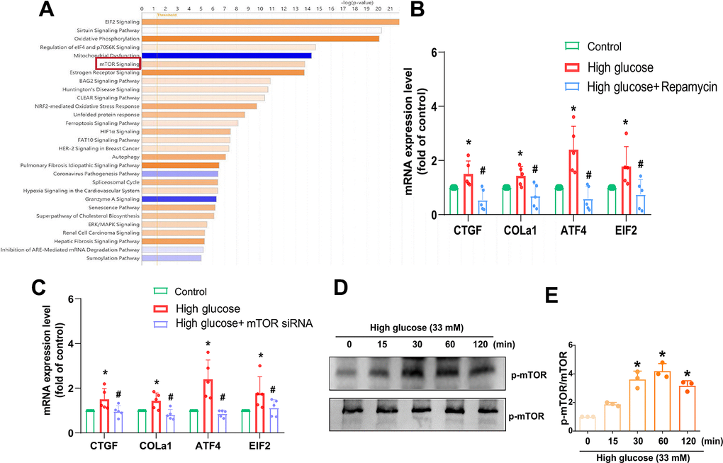
Figure 3.mTOR is regulated in HG-promoted fibrotic protein expression in HAFCs. (A) IPA pathway enrichment figure showing pathways in the GSE219145 dataset that significantly changed. (B, C) HAFCs were treated with mTOR inhibitor (rapamycin; 10 μM) or transfected with mTOR siRNA then treated with HG, and the indicated mRNA expression was examined by qPCR (n=5). (D) Cells were stimulated with HG, and the p-mTOR expression was examined by Western blot (n=3). (E) The densitometry analysis of (D) was quantified. * p < 0.05 versus the control group. # p < 0.05 versus the HG-treated group.