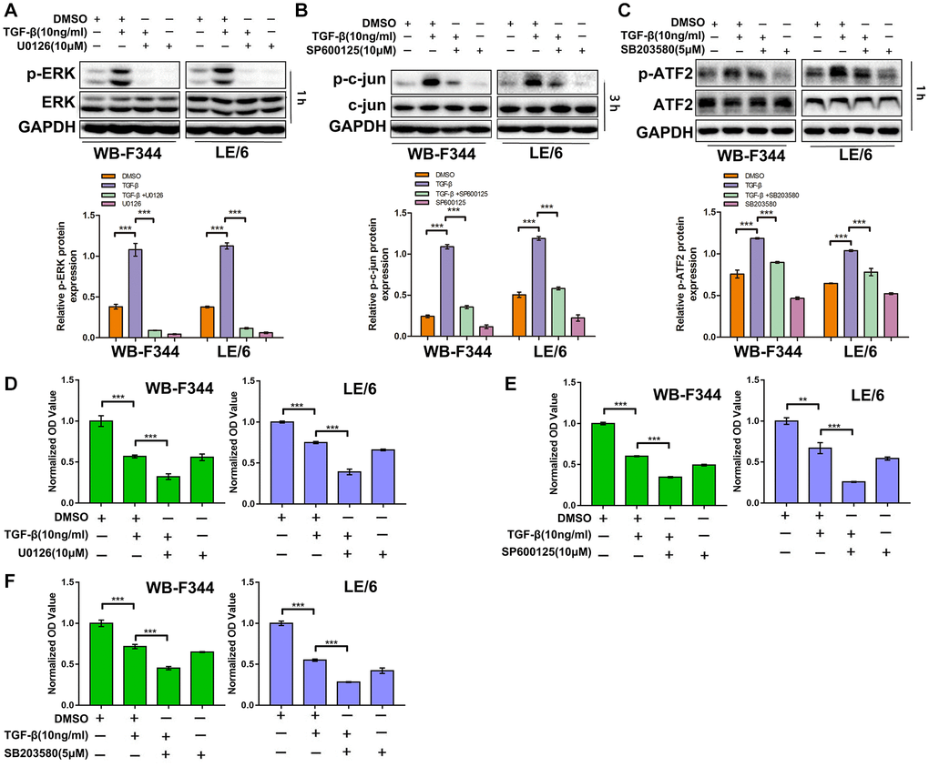
Figure 3.Suppression of TGF-β downstream of Erk, JNK or p38 MAPK signaling strengthens the TGF-β-induced cytostatic effects in LPCs. (A–C) WB-F344 and LE/6 cells were treated with TGF-β, U0126 (an Erk inhibitor), SP600125 (a JNK inhibitor), and/or SB203580 (a p38 MAPK inhibitor) as indicated, and Western blot analyses were carried out with antibodies against phospho-ERK and ERK (A), phospho-c-jun and c-jun (B), and phospho-ATF2 and ATF2 (C). GAPDH was used as a loading control. (D–F) WB-F344 and LE/6 cells were treated with TGF-β and/or the inhibitors as indicated for 3 days, after which CCK-8 analyses were performed. The normalized OD values of each group were compared, and the average OD values of three independent experiments are shown. One-way ANOVA was used for statistical analysis. The data are presented as the mean ± S.E.M. ***p < 0.001; ****p < 0.0001.