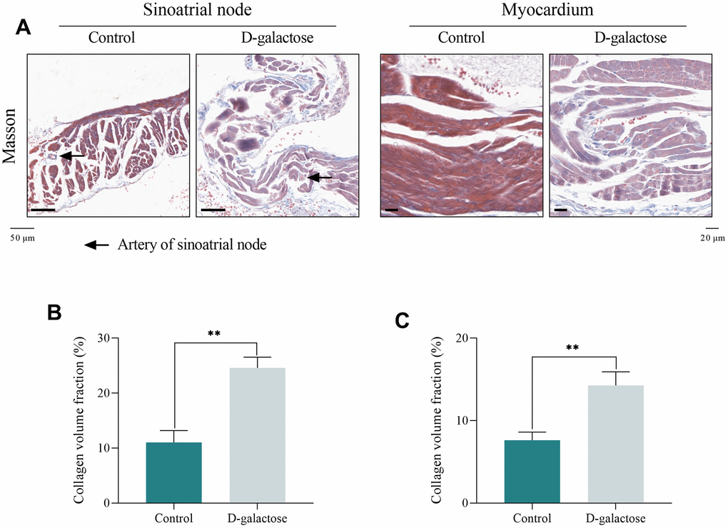
Figure 4.D-galactose causes fibrosis of sinoatrial node and myocardium. (A) Masson’s trichrome staining of sinoatrial node (Scale = 50 μm) and myocardium (Scale = 20 μm). (B) CVF of sinoatrial node. (C) CVF of myocardium. Black arrow represents the artery of sinoatrial node; ** represents P<0.01; Control: control group; D-galactose: D-galactose administered group.