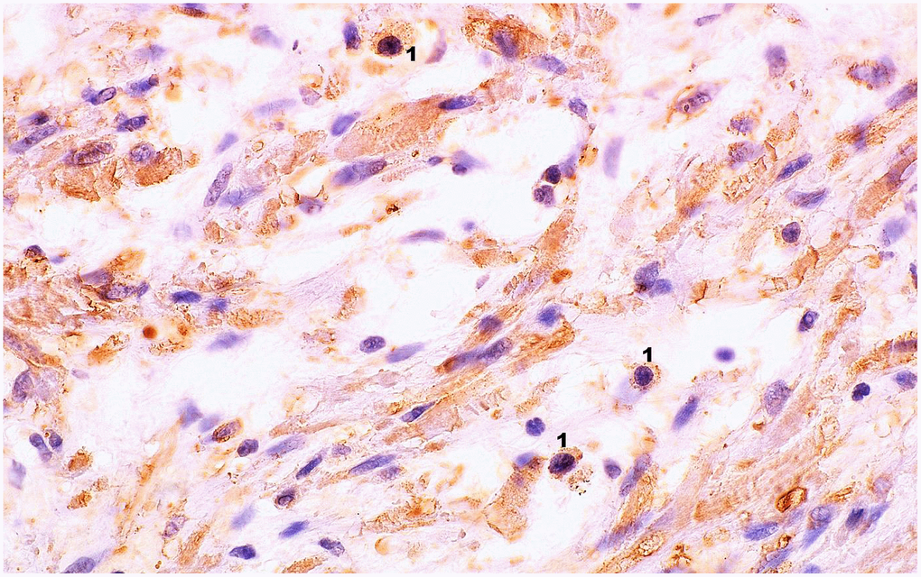
Figure 2.Microscopic image of the expression of the cytoplasmic immunohistochemical reaction to IL-6. Microscopic preparation of prostate stroma tissue with benign hyperplasia. Cytoplasmic immunohistochemistry showing IL-6 expression was stained brown (DAB +), 1 – lymphocytes, 600× magnification.