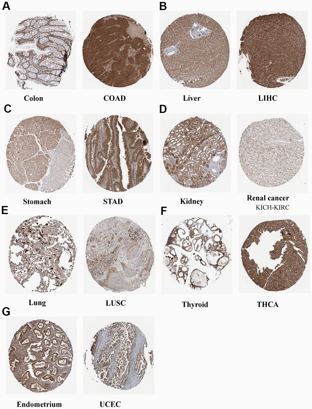
Figure 3.Representative IHC staining of N6AMT1 in eight normal (left) and tumor (right) tissues of the colon (A), liver (B), stomach (C), kidney (D), lung (E), thyroid (F) and endometrium (G).
Figure 3 — N6AMT1 is a novel potential diagnostic, prognostic and immunotherapy response biomarker in pan-cancer | Aging