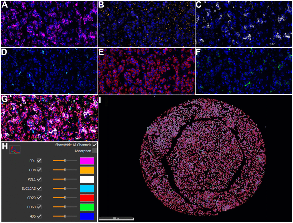
Figure 7.Multiplex immunohistochemistry profiling of SLC10A3 and immune markers in LGG. (A) PD1 (pink), (B) CD4 (yellow), (C) PD-L1 (white), (D) SLC10A3 (blue). (E) CD20 (red), (F) CD68 (green). (G) The merged image of seven markers. (H) Each marker stands for one special color. (I) Cell phenotype image constructed by the seven markers in the multiplex staining.