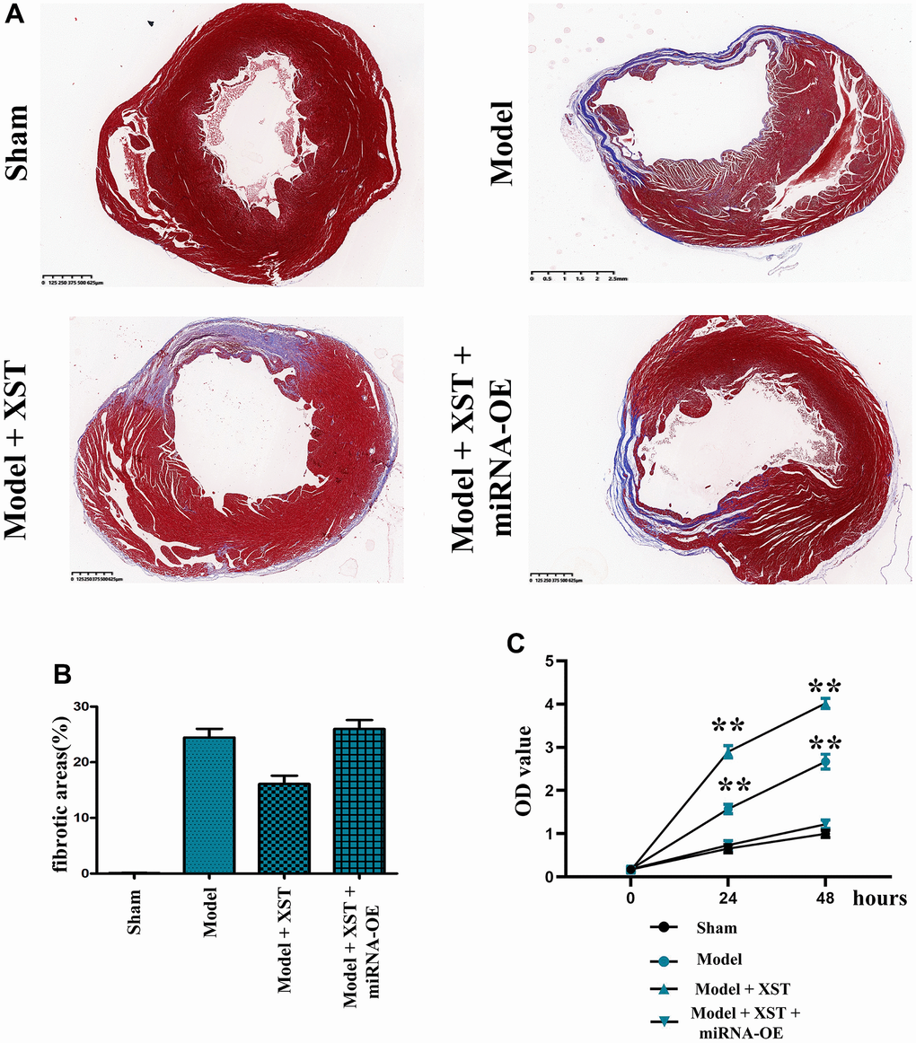
Figure 3.Changes of cardiac fibrosis in mice and cell proliferation capacity detected via MTT assay. (A) Masson staining of the mouse heart. (B) Statistical results of the proportion of cardiac fibrosis areas in mice. (C) The OD value detected by MTT.