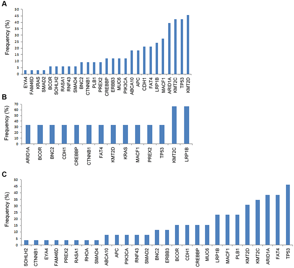
Figure 1.The frequency of genetic mutations according to the metastatic patterns. (A) Peritoneal metastasis, (B) hematogenous metastasis, and (C) distant lymphatic metastasis.
Figure 1 — Comparison of the mutation patterns between tumor tissue and cell-free DNA in stage IV gastric cancer | Aging