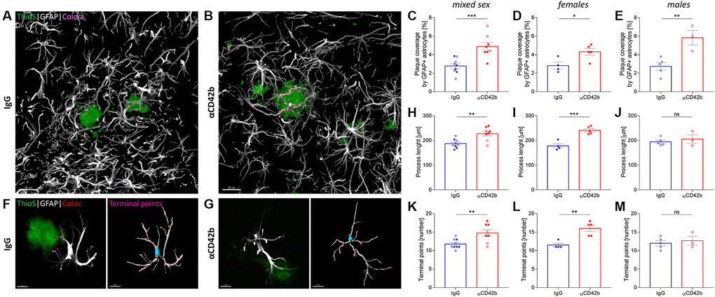
Figure 5.Platelet depletion increases astrocytic coverage of fibrillary amyloid plaques. (A, B) Brain tissue stained for amyloid plaques (green, thioflavin S) and astrocytes (white, GFAP) was used to analyze astrocyte-plaque interactions in the hippocampus. (C) Platelet-depleted mice showed increased overlap between astrocytic processes and amyloid plaques. (D, E) This effect was observed independent of sex. (F, G) Morphological analysis of plaque-associated astrocytes revealed increased (H–J) process length and (K–M) branching in platelet-depleted females but not in males. Data are shown as mean ± SEM. Statistical analysis was performed by unpaired Student’s t test (n = 8–9/group; “full forms” represent females and “empty forms” males). ***p < 0.001; **p < 0.01; *p < 0.05. Scale bar: 10 μm. Abbreviation: ThioS: thioflavin S.