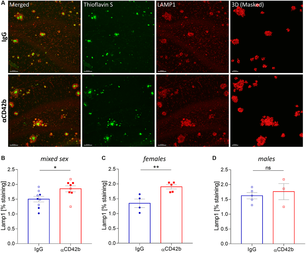
Figure 3.Platelet depletion increases neuritic dystrophy in the hippocampus of APP-PS1 females. (A) Brain tissue stained with thioflavin S (green, amyloid plaques) and LAMP1 (red, dystrophic neurites) was used for 3D modelling to quantify the volume of LAMP1+ clusters surrounding thioflavin S+ amyloid plaques. (B–D) Platelet depletion significantly increased plaque-associated dystrophic neurites in females but not in males. Data are shown as mean ± SEM. Statistical analysis was performed by unpaired Student’s t test (n = 8–9/ treatment; “full forms” represent females and “empty forms” males). **p < 0.01; *p < 0.05. Scale bar: 80 μm and 50 μm (3D mask).