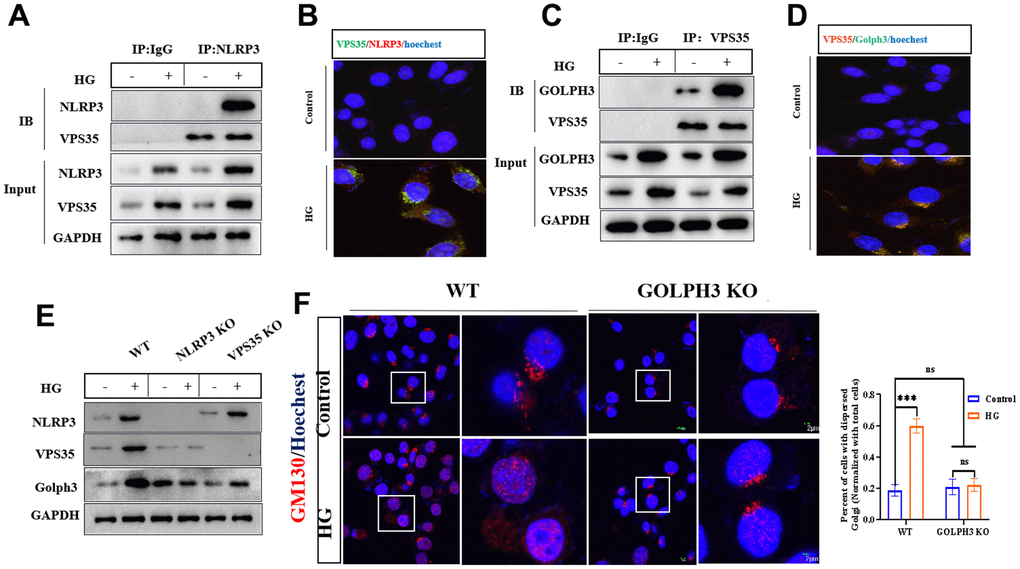
Figure 2.HG promotes the activation of NLRP3/VPS35/Golph3 pathway. (A) The interaction of NLRP3 and VPS35 was determined by co-immunoprecipitation experiment. Lysates from BV2 microglial cells were immunoprecipitated with a NLRP3 antibody or rabbit IgG and immunoblotted with VPS35 and NLRP3 antibody. (B) The colocalization of NLRP3 and VPS35 was tested by immunofluorescence method. Bar = 2 μm. (C) The interaction of VPS35 and Golph3 was determined by co-immunoprecipitation experiment. Lysates from BV2 microglial cells were immunoprecipitated with a VPS35 antibody or rabbit IgG and immunoblotted with VPS35 and Golph3 antibody. (D) The colocalization of VPS35 and Golph3 was tested by immunofluorescence method. Bar = 2 μm. (E) Western blot analysis of NLRP3, VPS35, and Golph3 in WT, NLRP KO and VPS35 KO BV2 cells treated with or without HG. GAPDH was used as an internal control for normalization. (F) Immunofluorescence detection of GM130 in WT and Golph3 KO BV2 cells, the percent of cells with dispersed Golgi apparatus normalized with total cells. Bar = 2 μm. Data represent means ± SEM of 3 independent experiments. * p ≤ 0.05, ** p ≤ 0.01, and *** p ≤ 0.001 according to two-way ANOVA with Bonferroni's post hoc test.