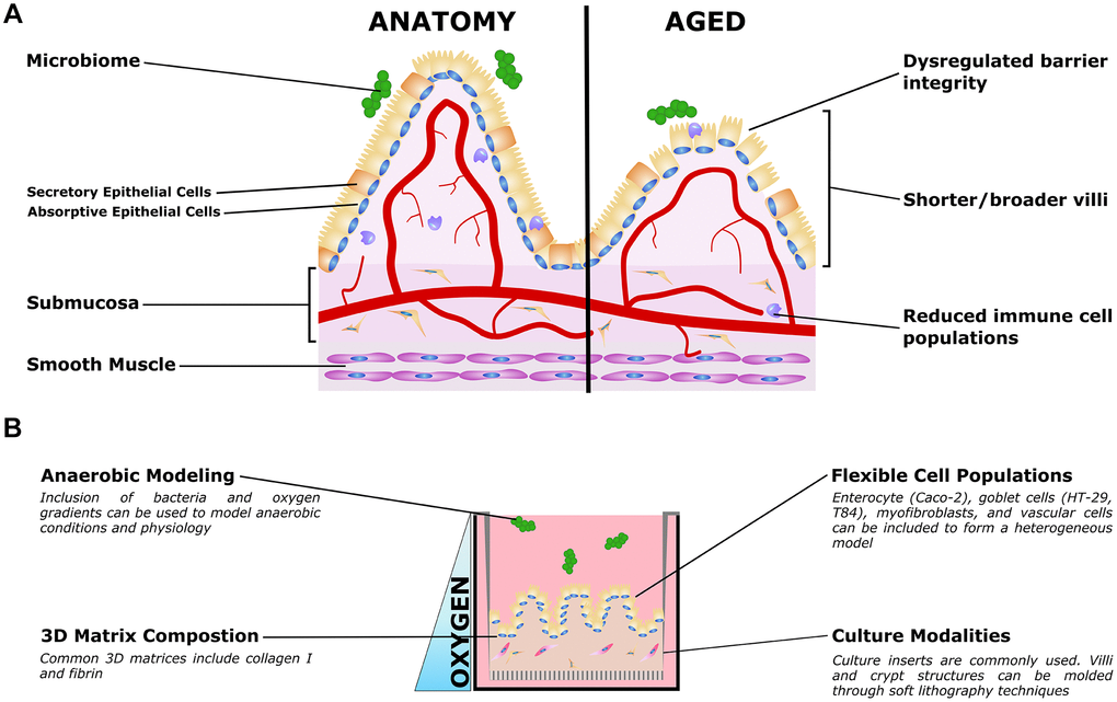
Figure 2.Organotypic models of gut aging. (A) Simplified gut anatomy and aging, focusing on the most commonly modeled components. A mixed epithelial population, described in the text, forms a simple cuboidal epithelial layer with both secretory and absorptive epithelium. A layer of mucus inside the gut lumen supports the host/microbiome interaction. The stroma underneath the epithelium, the submucosa, is host to nerves (not shown) blood vessels, fibroblasts, and immune cells important for gut function. Smooth muscle is required for gut peristalsis. In aging, the macrostructure of villi degrades, with villi becoming shorter and broader. Immune cell populations are disrupted, and reduced epithelial barrier integrity can lead to increased microbial infiltration into the submucosa and vasculature. (B) Organotypic models of the gut typically only model a small subset of these features, and are typically adapted to aspects that are relevant to specific questions. For example, epithelial and immune populations may be co-cultured to study intercellular interactions in a simple format. To study the influence of villous structures, soft lithography can be used to recreate the villi/crypt geometry. Microbiome co-cultures can be included, and microfluidic organ-on-a-chip models have been used to mimic the oxygen gradient from the vascularized submucosa to the anaerobic lumen.