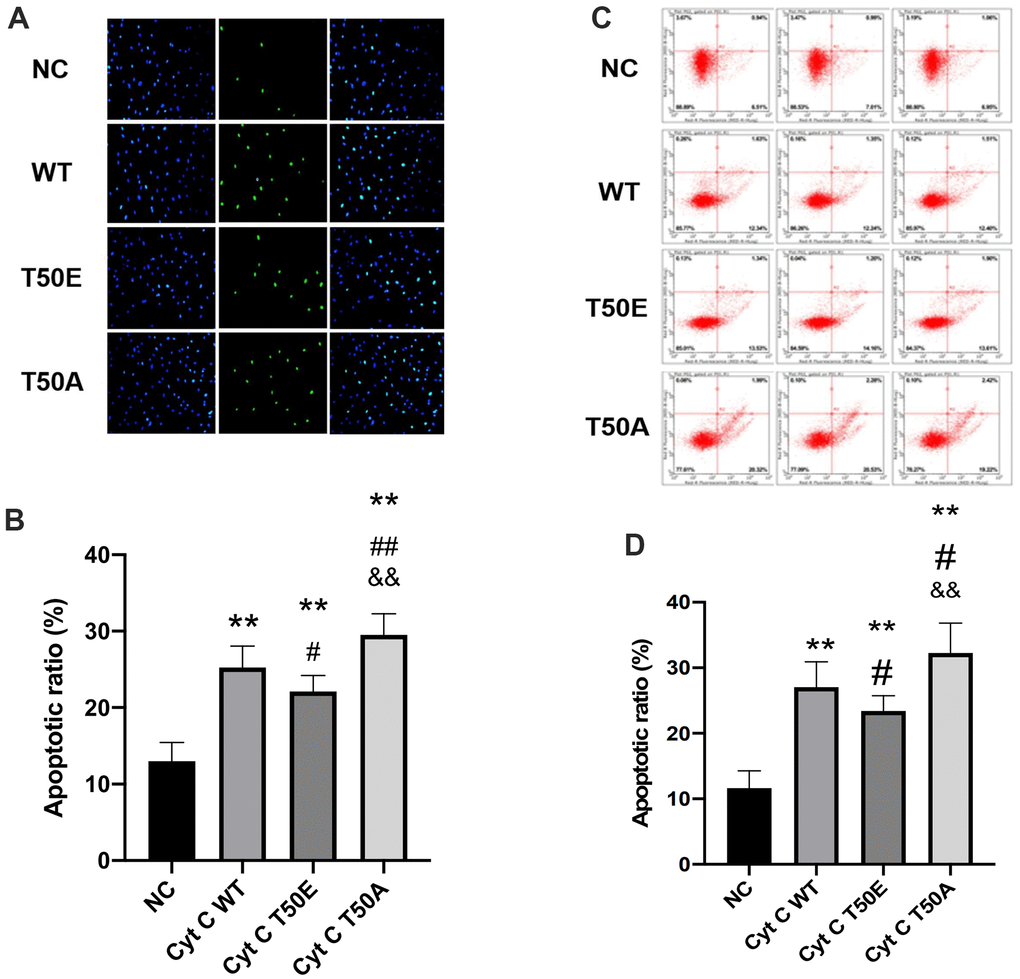
Figure 4.Cytc T50 phosphorylation suppresses H/R induced apoptosis. (A) Representative photomicrographs of in situ detection of cardiomyocytes DNA fragments from AC16 cells subjected to H/R. Cardiomyocytes were stained with DAPI (blue) and TUNEL (green). (B) TUNEL-positive nuclei were summarized in a graph and expressed as a percentage of all cardiomyocytes subjected to H/R. (C) Apoptosis in AC 16 cells were analyzed by flow cytometry. (D) Quantitation of apoptosis data in flow cytometry. n=3 in each group. Data expressed as mean±SD. ** P < .01 vs. NC, # P < .05 vs. WT, ## P < .01 vs. WT, && P < .01 vs. T50E.