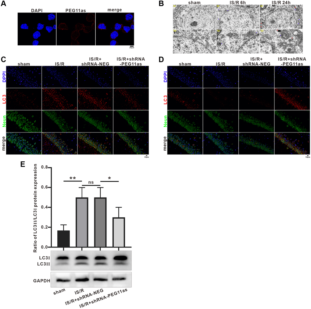
Figure 3.PEG11as silencing inhibited neuronal autophagy and apoptosis. (A) A FISH assay showed the location of PEG11as in mouse primary neurons. Green, PEG11as; Blue, DAPI. (B) Transmission electron microscopy was applied to observe the ultrastructural features in hippocampal of tMCAO/R mice. Blue arrow indicated autophagosomes and yellow arrow represented lysosomes. (C, D) Representative double immunofluorescent staining for NeuN and MAP1LC3B (C) and SQSTM1/p62 (D) in ischemic hemispheres transfected with shRNA-PEG11as 14 days and treated with tMCAO/R. (E) Representative pictures and statistical chart for the western blot of MAP1LC3 staining. n = 3. One-way ANOVA followed by the Tukey’s post-hoc-test was used, data are statistically different from each other with *P < 0.05, **P < 0.01 and ***P < 0.001. Abbreviation: ns: no statistically different vs. IS/R group.