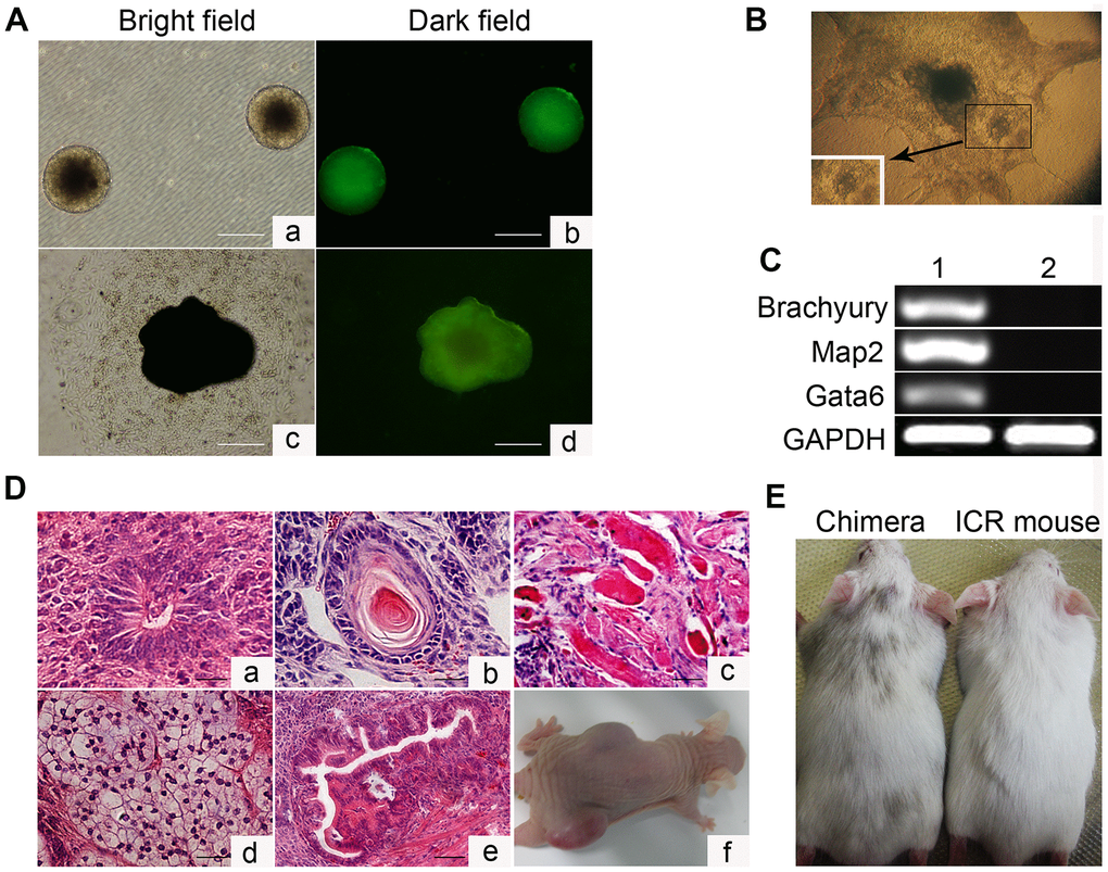
Figure 2.In vitro embryoid body-mediated differentiation and in vivo developmental pluripotency of Oct4-EGFP miPSCs. (A) In vitro embryoid body (EB) formation (a, b) and differentiation (c, d). (B) In vitro EB differentiation into myocardium cells. (C) RT-PCR analyses of various differentiation markers for the following three germ layers in EB. Brachyury (a marker of mesoderm), microtubule associated protein 2 (Map2, ectoderm), and GATA-binding factor 6 (Gata6, endoderm). (D) Various tissues present in teratomas derived from Oct4-EGFP miPSCs. (E) Chimeric mouse generated by Oct4-EGFP miPSCs. Scale bar: (A, D) 50μm.