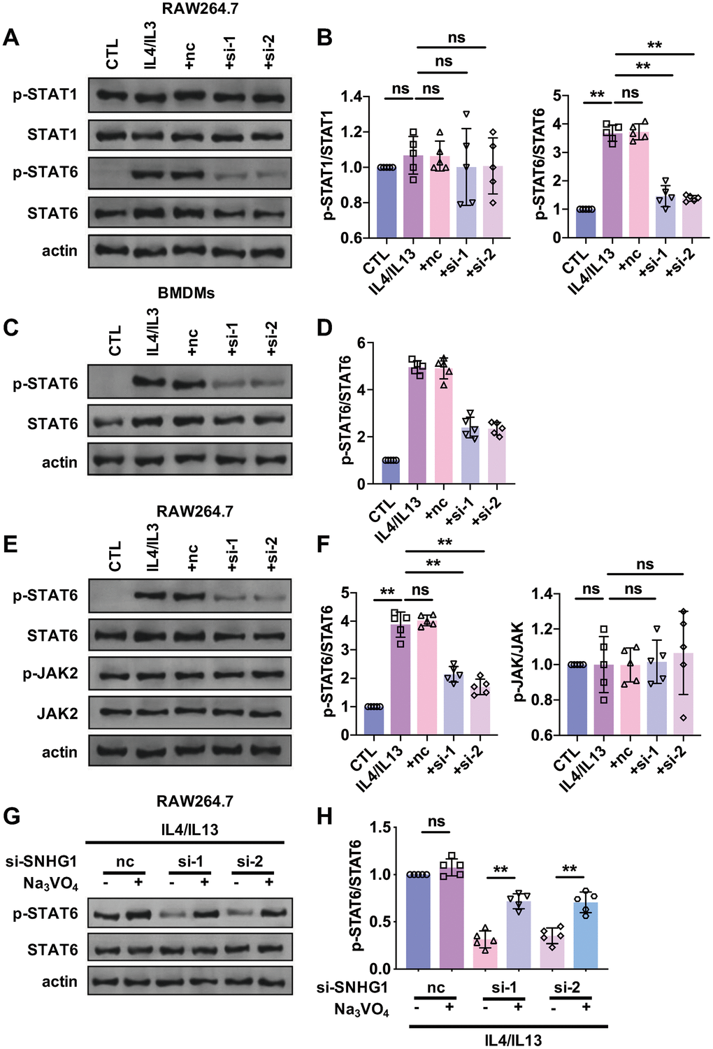
Figure 4.STAT6 was involved in the regulation of M2 macrophage polarization mediated by lncRNA-SNHG1. (A, B) Western blot was performed to detect the protein levels of STAT1, p-STAT1, STAT6, and p-STAT6. RAW264.7 cells were transfected with two siRNAs of lncRNA-SNHG1 and negative control siRNA for 48h and treated with IL4/IL13 or LPS/INFγ for 72h. one-way ANOVA was used for the statistical analysis. n=5 independent cell cultures. The bar indicates the SD values. **P<0.01. (C, D) Western blot was performed to detect the protein levels of STAT6 and p-STAT6. BMDMs were transfected with two siRNAs of lncRNA-SNHG1 and negative control siRNA for 48h and treated with IL4/IL13 or LPS/INFγ for 72h. one-way ANOVA was used for the statistical analysis. n=5 independent cell cultures. The bar indicates the SD values. **P<0.01. (E, F) Western blot was performed to detect the protein levels of STAT6, p-STAT6, JAK, and p-JAK. RAW264.7 cells were transfected with two siRNAs of lncRNA-SNHG1 and negative control siRNA for 48h and treated with IL4/IL13 or LPS/INFγ for 72h. one-way ANOVA was used for the statistical analysis. n=5 independent cell cultures. The bar indicates the SD values. **P<0.01. (G, H) Western blot was performed to detect the protein levels of STAT6 and p-STAT6. RAW264.7 cells were transfected with two siRNAs of lncRNA-SNHG1 and negative control siRNA for 48h and treated with IL4/IL13 or LPS/INFγ for 72h. one-way ANOVA was used for the statistical analysis. n=5 independent cell cultures. The bar indicates the SD values. **P<0.01.