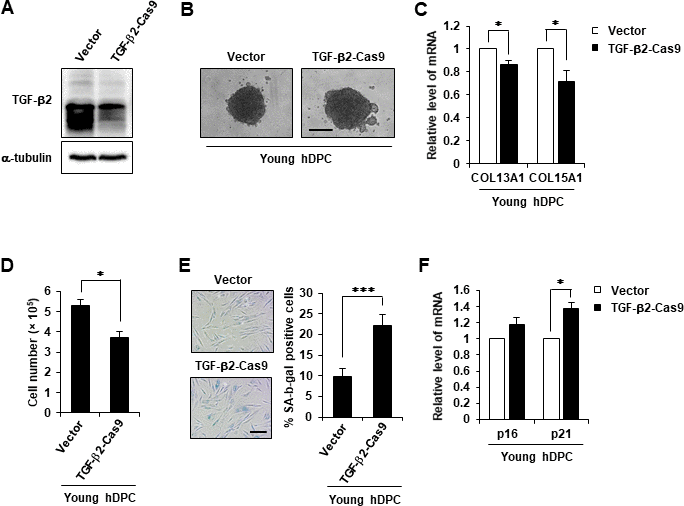
Figure 6.TGF-β2 KO induced cellular senescence. (A) Western blot analysis of TGF-β2 in TGF-β2 knockout hDPCs (P5). (B) Representative image of hDPC spheroids transfected with control or TGF-β2 CRISPR/Cas9 KO plasmid for 48 h. (C) COL13A1 and COL15A1 mRNA expression in 3D spheroids of CRISPR/Cas9 KO plasmid-transfected hDPC. (D) Cell numbers after control or TGF-β2 CRISPR/Cas9 KO plasmid transfection for 48 h. (E) Quantification and representative images of SA-β-gal-positive cells in control and TGF-β2 CRISPR/Cas9 KO plasmid-treated hDPCs. Scale bar, 200 μm. (F) p16 and p21 mRNA expression by qRT-PCR after CRISPR/Cas9 KO plasmid transfection for 48 h. All quantitative data are shown as the mean ± SD of three independent experiments. *p < 0.05; ***p < 0.005. Asterisk indicates Student's t-test.