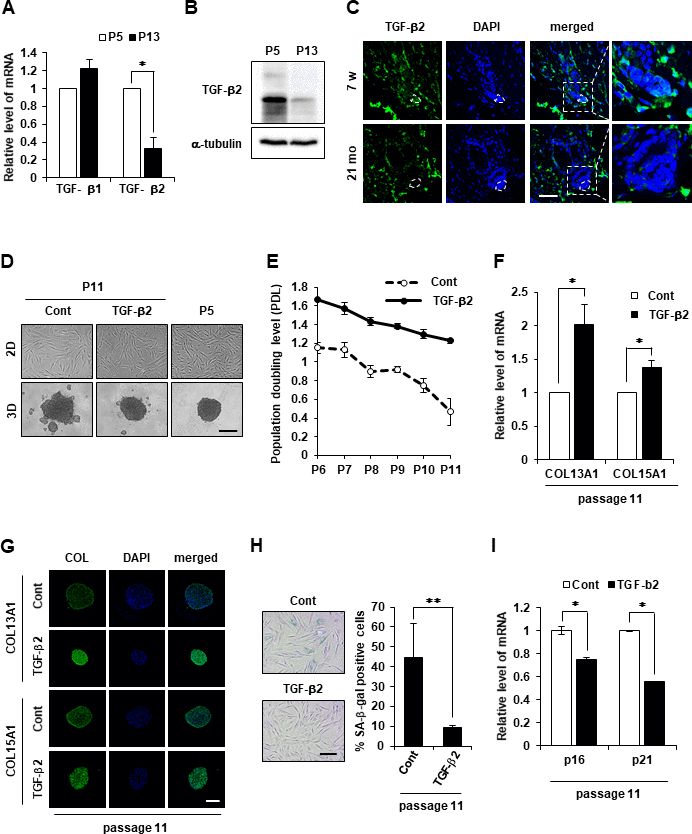
Figure 5.TGF-β2 supplement maintains cell aggregative behavior and prevents senescence in DPCs. (A) TGF-β1 and TGF-β2 mRNA expression was determined by qRT-PCR in P5 and P13 DPC spheroids. (B) TGF-β2 expression was determined by western blotting in P5 and P13 DPC spheroids. (C) Skin biopsies of normal C57BL/6 mice were collected at the indicated age and processed for paraffin sections. TGF-β2 expression was visualized by immunofluorescence staining and counterstained with DAPI for nuclei. The DP was circled by white dashed lines in each HF. Scale bar, 50 μm. (D) Representative image of 2D and 3D spheroids of hDPCs cultured in the absence or presence of TGF-β2. Cells were sustained with TGF-β2 (50 ng/ml) in the culture medium from P5 to P11, and the total cell lysates from P11 were used. Scale bar, 200 μm. (E) Cell duplication level between consecutive passages of DPCs in the presence or absence of TGF-β2 (50 ng/ml) was investigated by cell counting. To calculate the duplication level of each passage, the harvested cell number was normalized to the seeding cell number at a 2-day interval. Experiments were carried out in triplicates. COL13A1 and COL15A1 mRNA expression by qRT-PCR (F) and immunofluorescence image (G) in 3D spheroids at P11 of hDPCs cultured in the absence or presence of TGF-β2. Scale bar, 200 μm. (H) Quantification and representative images of SA-β-gal-positive cells in P11 of hDPCs cultured in the absence or presence of TGF-β2. (I) p16 and p21 mRNA expression by qRT-PCR in P11 of hDPCs cultured in the absence or presence of TGF-β2. All quantitative data are shown as the mean ± SD of three independent experiments. *p < 0.05; **p < 0.01. Asterisk indicates Student's t-test.