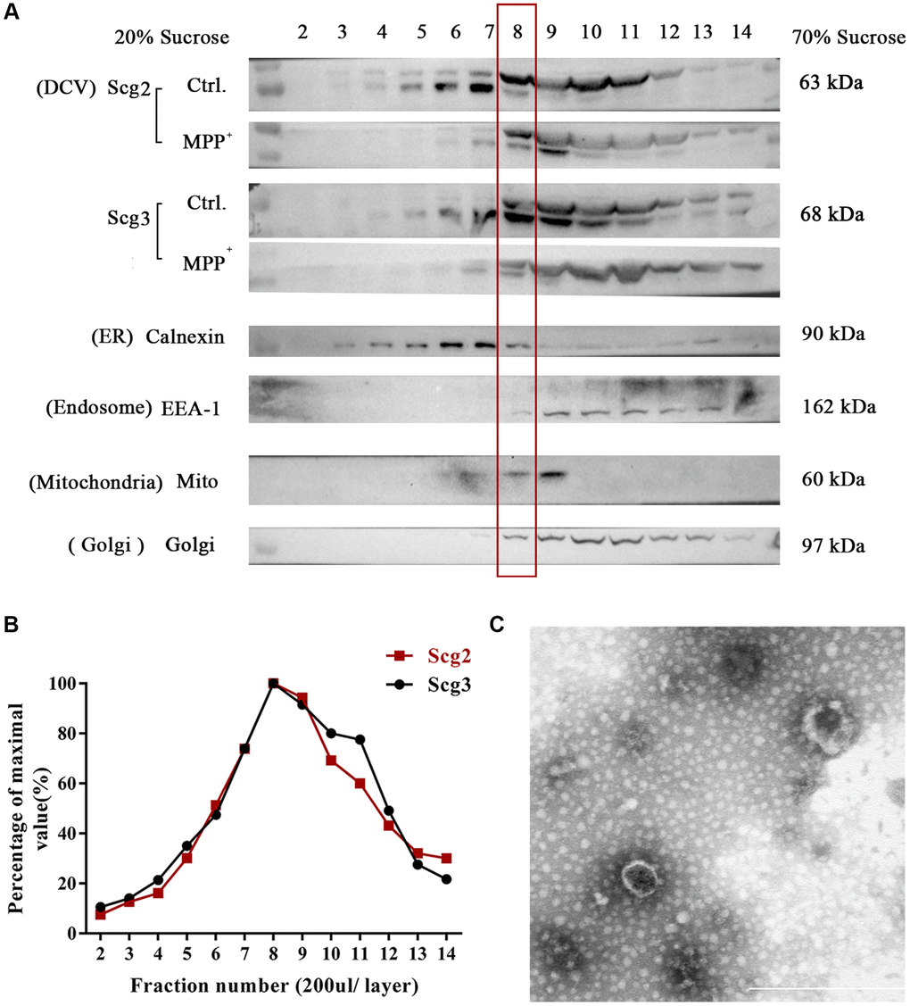
Figure 4.Fractionation of components in SH-sy5y cells via sucrose density gradient centrifugation. (A) The extract of SH-sy5Y was fractionated into 17 layers via sucrose density gradient centrifugation. And levels of multiple organelles, secretory granules (Scg2 and Scg3), ER (Calnexin), Golgi (Golgi 97), endosome (EEA-1), and mitochondria (Mito) were measured by immunoblotting for each fraction (layer 2-14). (B) The protein levels of Scg2 and Scg3 in fractions 2–14 were quantified and showed in a line graph. And layer 8–9 are peak fractions for both proteins. (C) Transmission electron microscopy of the purified secretory granules in the 8th fraction of SH-sy5y cells via sucrose density gradient centrifugation. Scale bar = 500 nm.