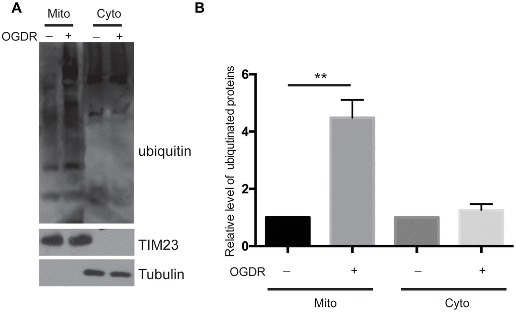
Figure 1.Ubiquitination of mitochondrial and cytoplasmic proteins in the early stage of reperfusion after 4 h OGD. (A) Western blot using an antibody against ubiquitin was performed to examine the ubiquitination of mitochondrial (Mito) and cytoplasmic (Cyto) proteins after 45 min reperfusion following 4 h OGD in SK-N-BE(2) cells. (B) Quantitation (Mean ± SEM) of A from three independent experiments. There was a significant increase in the ubiquitination of mitochondrial proteins during early reperfusion after 4 h of OGD, but not in the ubiquitination of the cytoplasmic proteins.