Submit an Article
Navigate
Home
Editorial Board
Editorial Policies
Current Volume
Archive
Scientific Integrity
Publication Ethics Statements
Interviews with Outstanding Authors
Newsroom
Sponsored Conferences
Podcast
Contact
Special Collections
Submit an Article
Online ISSN: 1945-4589
Research Paper
|
Volume 12, Issue 24
|
pp. 24709–24720
Post-transplant colitis after kidney transplantation: clinical, endoscopic and histological features
Back to article
Figure 3
(3 of 6)
−
100%
+
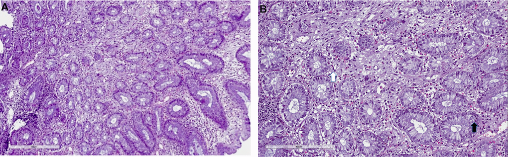
Figure 3.
Mycophenolate mofetil colitis.
Histological examination. Biopsy of right colon: (
A
) severe eosinophils, lymphocytes and plasma cells infiltrate (10x), with (
B
) severe cryptitis (20x, white arrow) and cell apoptosis (black arrow).