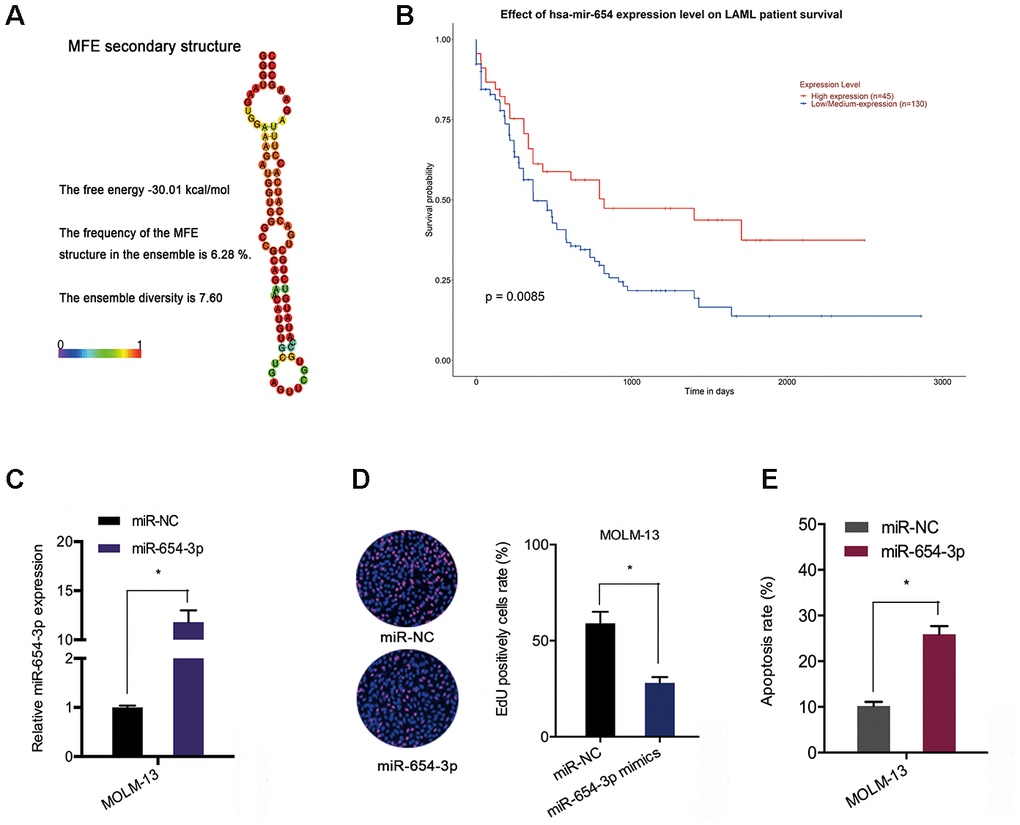
Figure 5.MiR-654-3p reduced AML cell progression. (A) The secondary structure of miR-654-3p. (B) TCGA database showed that reduced miR-654-3p levels was related to poor OS in AML patients. (C) The transfection efficiency of miR-654-3p mimics in AML cells. (D, E) MiR-654-3p mimics decreased AML cell growth and stimulated cell apoptosis in vitro. *P<0.05.