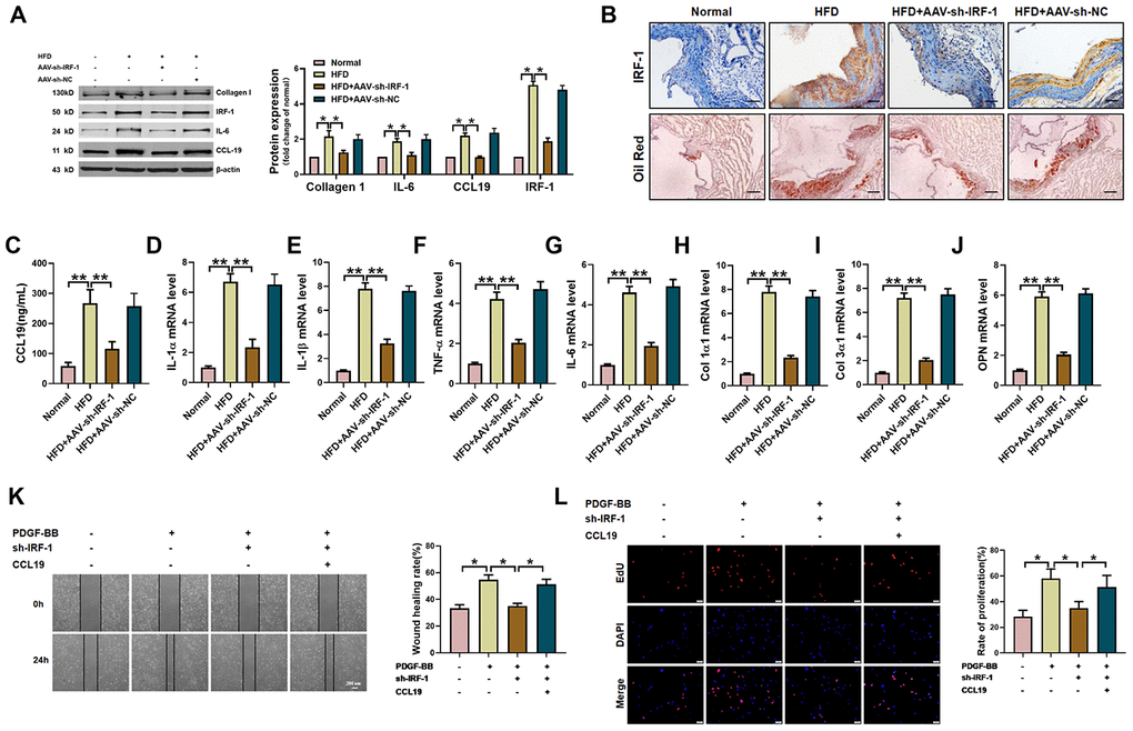
Figure 6.IRF-1 knockdown inhibits AS of mice and proliferation and migration of VSMCS. (A) Western blot was used to analyze the protein level of IL-6, Collagen 1, CCL19 and IRF-1 in AS mice and AAV-sh-IRF-1 treated mice. β-actin was used as an internal control. n=4 (B) Representative images of IHC staining and oil red staining in AS model mice aorta tissues and normal aorta tissues. Bar=50 μm. Knockdown IRF-1 reversed HFD induced mRNA expression of CCL19 (C), inflammatory cytokine IL1-α (D), IL-1β (E), TNF-α (F), IL-6 (G) and the deposition of ECM collagen1 (H), collagen3 (I) and OPN (J). n=56. In vitro, sh-IRF-1 led migration (K) and proliferation (L) of VSMCS. n=6. *P<0.05; **P<0.01.