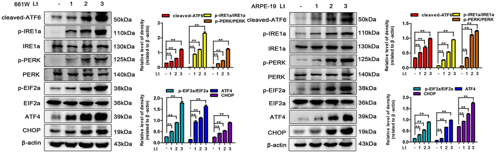
Figure 3.Light exposure induces ER stress in photoreceptors and RPEs. 661W cells and ARPE-19 cells were cultured in a dark condition or exposed to 1500 Lux light for 1–3 days after which the levels of ER stress markers were determined by western blotting. β-actin was referenced as an internal control. Three independent experiments are conducted two weeks apart. The results are presented as the mean± SEM. n (per group) =3, **P < 0.01.