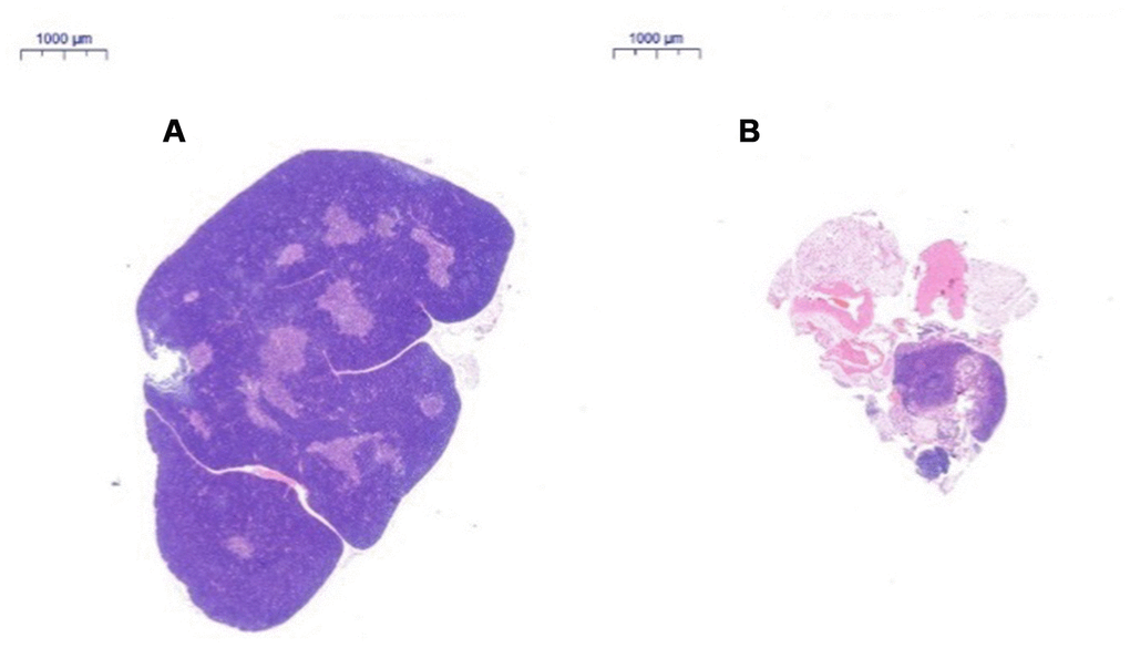
Figure 3.Changes in the thymus tissue structure in C57 mice of different ages. Note: (A) shows the thymus from a 2-month-old C57 mouse (20×); (B) shows the thymus from an 18-month-old C57 mouse (20×). HE staining of the thymus in 18-month-old C57 mice: the volume of the thymus was reduced, and the boundary of the cortex and medulla was not clear; most of the thymus tissue had been replaced by adipose tissue.