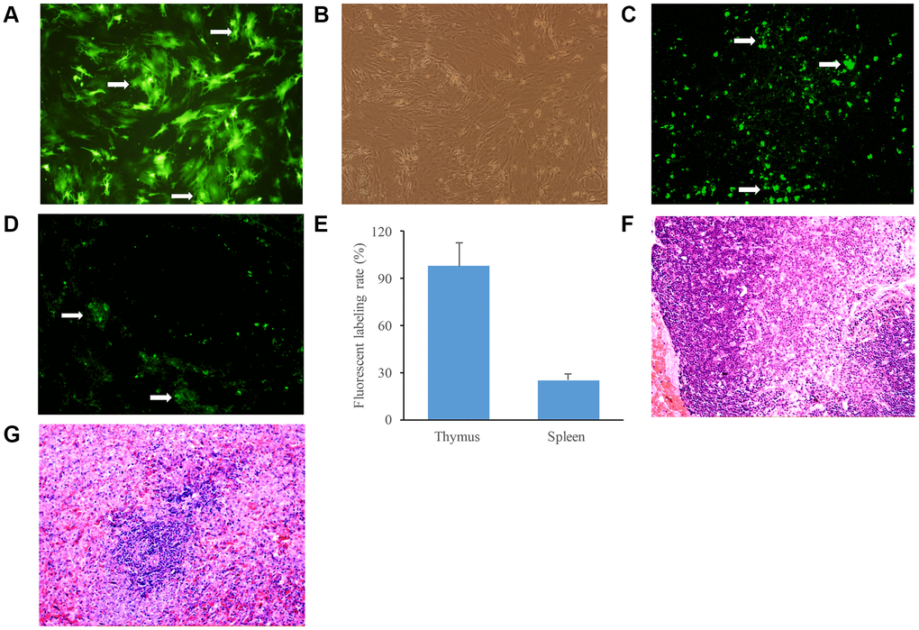
Figure 2.Identification of GFP labeled BMSCs in the thymus and spleen tissues. (A) BMSCs were observed after transfection with GFP for 24 h; (B) BMSCs in the same field were observed by ordinary inverted microscope; (C) GFP labeled BMSCs were observed in the thymus tissues of aging rats after infusion with BMSCs; (D) GFP labeled BMSCs were observed in the spleen tissues of aging rats after infusion with BMSCs; (E) The fluorescent labeling rate was measured after infusion with BMSCs; (F) The thymus tissues of aging rats were stained by HE staining; (G) The spleen tissues of aging rats were stained by HE staining. White arrows indicate green fluorescent cells.