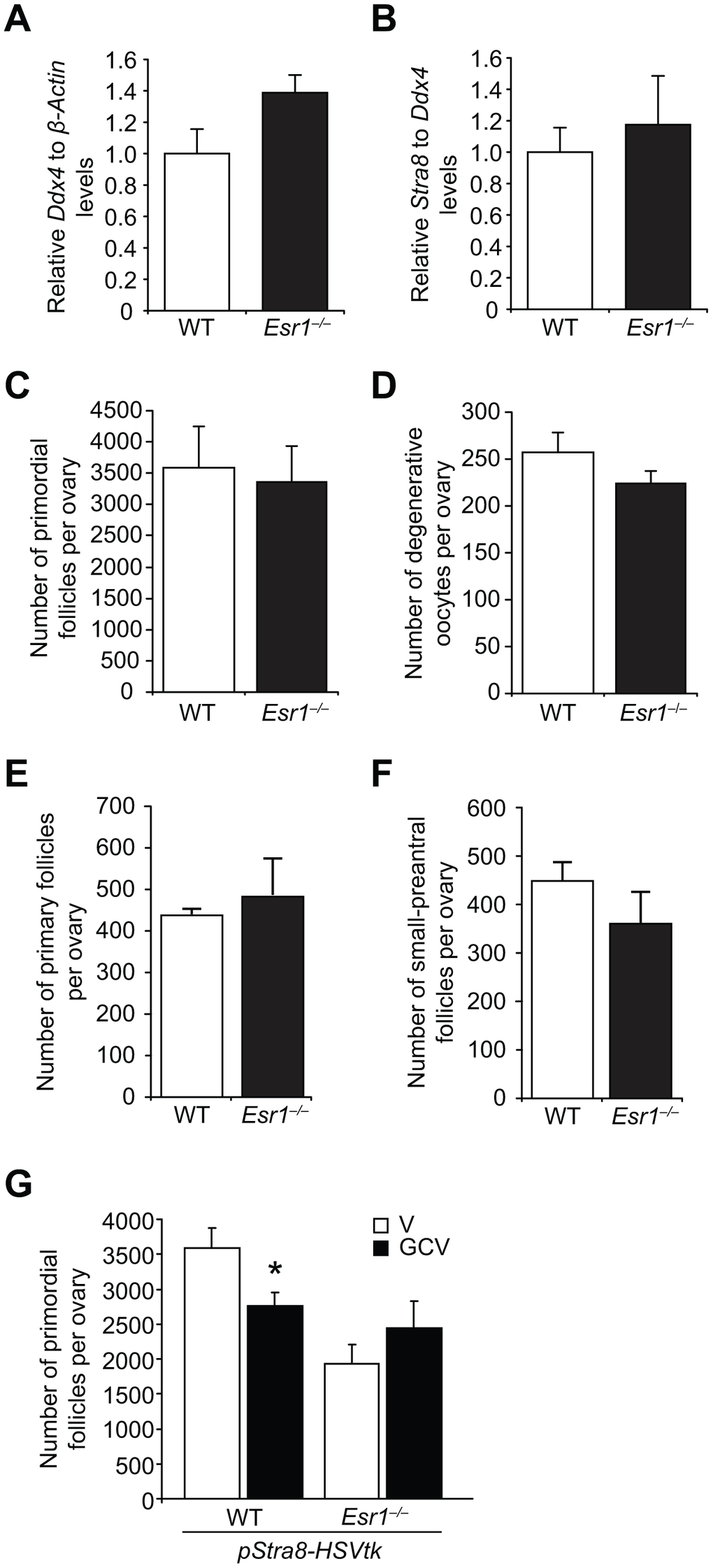
Figure 6.Esr1-null female mice exhibit impaired postnatal oocyte renewal. (A, B) Expression of Ddx4 (A; normalized to β-actin, indicative of the relative numbers of germ cells in whole gonads) and Stra8 (B; normalized to Ddx4, indicative of the relative level of Stra8 activation across the total germ cell pool) in e13.5 ovaries collected from WT and Esr1–/– female fetuses (mean ± SEM, n = 10 timed-pregnant female mice, with fetal ovaries of each genotype collected from each timed-pregnant dam serving as an independent replicate). (C) Primordial follicle numbers in ovaries of neonatal (5-day-old) Esr1–/– mice compared to WT littermates (mean ± SEM, n = 6 mice per group). (D) Numbers of degenerative oocytes in ovaries of young adult Esr1–/– mice compared to WT littermates (mean ± SEM, n = 4–6 mice per group). (E, F) Numbers of recently growth-activated (primary; E) and early growing (small-preantral; F) immature follicles in ovaries of Esr1–/– mice, compared to WT littermates, at 2 months of age (mean ± SEM, n = 4–6 mice per group). (G) Primordial follicle numbers in ovaries of young adult pStra8-HSVtk;WT and pStra8-HSVtk;Esr1–/– mice treated with vehicle (V) or GCV (10 mg/kg) for 21 days (mean ± SEM, n = 5–6 mice per group; *P<0.05).