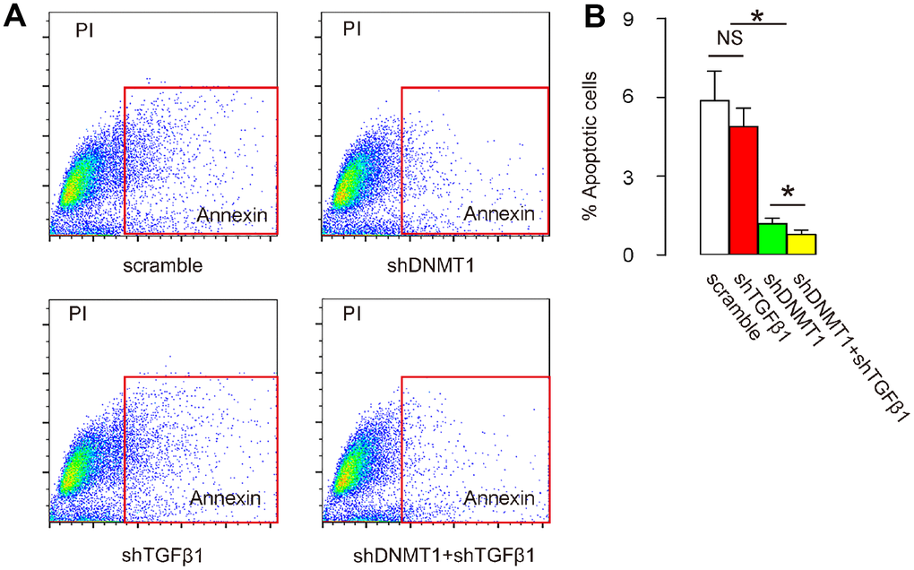
Figure 5.Co-application of shDNMT1 and shTGFβ1 reduces cell apoptosis in LDD tissue. (A, B) Cells isolated from mouse vertebral pulp and annulus fibrosus were dissociated into single cell populations for a flow-cytometry-based apoptosis assay, shown by representative flow charts (A) and by quantification (B). *p<0.05. NS: non-significant. N=7.