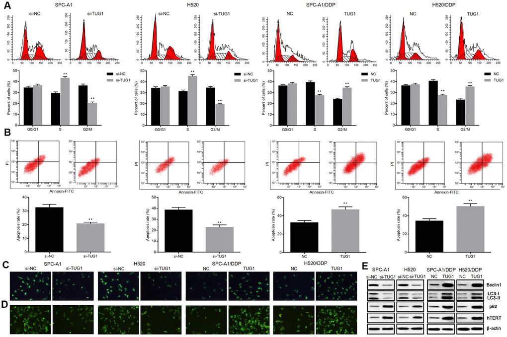
Figure 3.Overexpression of TUG1 enhanced the apoptosis, autophagy and senescence of NSCLC cells in response to DDP. (A) the effect of TUG1 on the cell cycle distribution of NSCLC cells; (B) the effect of TUG1 on apoptosis of NSCLC cells; (C) GFP-LC fusion protein tagging detection of autophagosome formation; (D) MDC staining to detect formation of autophagic vacuoles; (E) expression of autophagy-related proteins and cell senescence-related protein hTERT determined by Western blot analysis, normalized to β-actin. ** p < 0.01 versus the control group.