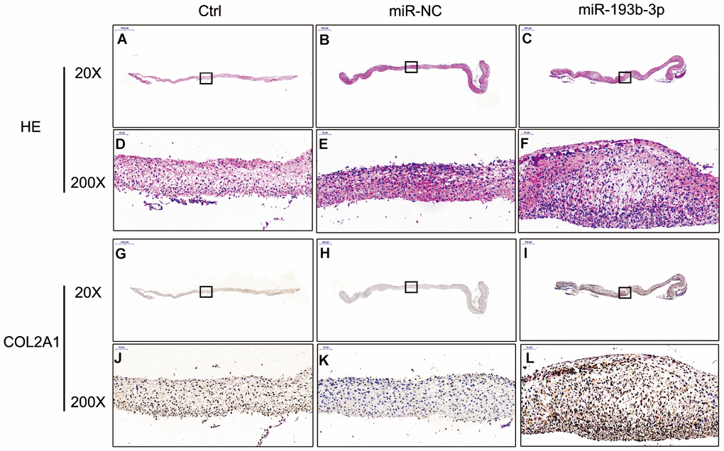
Figure 3.In vitro H&E staining and immunohistochemical assay of the chondrocyte sheets in different groups (n=3). (A–C) H&E staining magnification 20 times (bar 500 μm). (D–F) Zoom square magnification x200; bar 50 μm. (G–I) Immunohistochemical assay magnification 20 times (bar 500 μm). (J–L) Zoom square magnification x200; bar 50 μm.