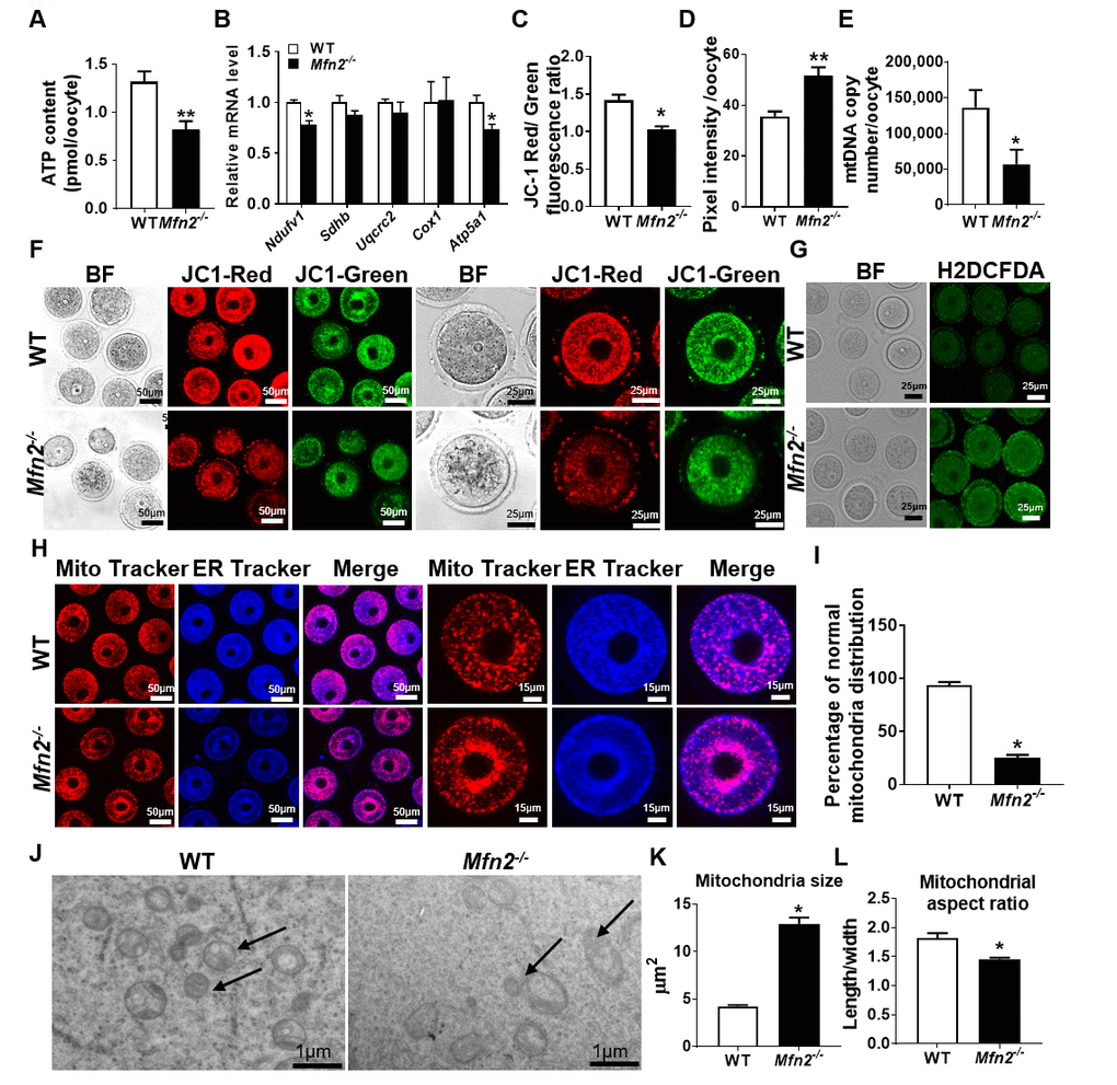
Figure 2.Mitochondrial function is impaired in Mfn2-/- oocytes. (A) ATP measurement in Mfn2-/- and WT mice GV stage oocytes. (B) mRNA expression of respiratory chain genes was assessed using qRT-PCR in GV stage oocytes collected from Mfn2-/- and WT mice. (C, F) Representative fluorescent micrographs of GV stage oocytes stained by mitochondria JC-1. Red fluorescence represents J-aggregate while green fluorescence represents JC-1 monomer. Mitochondrial membrane potential is indicated by the red/green fluorescence intensity ratio. (D, G) Fluorescence intensity of Carboxy-H2DCFDA was used to measure ROS levels after treatment with H2O2. (E) mtDNA copy number was determined by qRT-PCR in GV stage oocytes collected from Mfn2-/- and WT mice. (H) Mitochondria and ER were labeled by immunostaining with MitoTracker (red) and ER-Tracker (blue). (I) The percentages of oocytes with normal distribution of mitochondria in the Mfn2-/- and WT mice. (J) Representative electron microscopic graphs of oocytes from 8-week-old Mfn2-/- and WT mice (n=3 ovary from different mice assessed in each group). Arrows show mitochondria. (K, L) Mitochondrial size and aspect ratio in Mfn2-/- and WT oocytes. Data presented as mean ± SEM. *p < 0.05, **p < 0.01 vs. WT from t-test. ATP: Adenosine triphosphate. Ndufv1: NADH dehydrogenase (ubiquinone) flavoprotein 1; Sdhb: succinate dehydrogenase complex iron sulfur subunit B; Uqcrc2: ubiquinol cytochrome c reductase core protein 2; Cox1: cytochrome c oxidase subunit I; Atp5a1: ATP synthase, H+ transporting, mitochondrial F1 complex, alpha subunit 1.