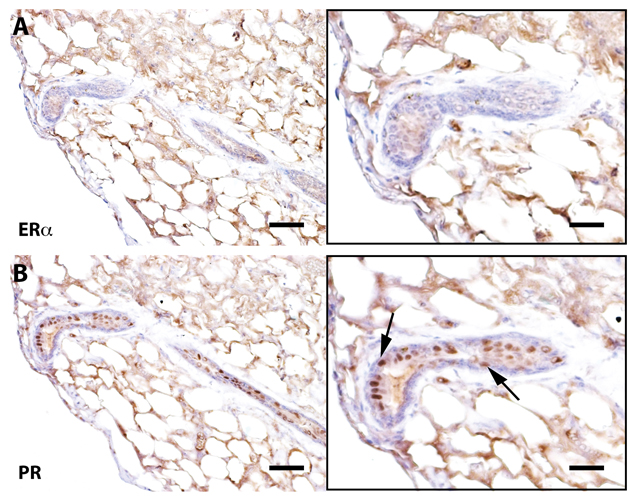
Figure 3.Senescent outgrowths contain PR+ cells but lack ER-α+ signaling cellsImmunohistochemical staining of a cross-section from Wap-Cre/TGF-β1+ outgrowths stained for ER-α and progesterone receptor (PR). Arrows denote PR positive cells. Scale bars: 40 μm left panels, 80μm right panels.