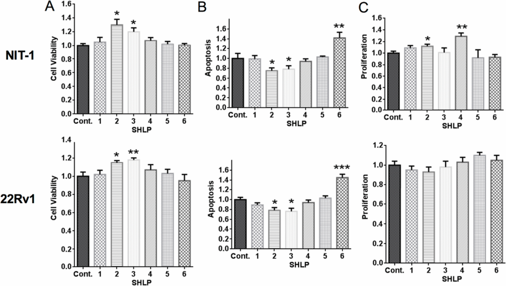
Figure 2.Effect of SHLPs on cell growth and deathNIT-1 β and 22Rv1 cells were cultured in serum-free (SF) media with 100 nM SHLP or control peptides and assessed for (A) cell viability (using the MTS assay) after 72 h; (B) apoptosis after 24 h; and (C) cell proliferation by BrdU incorporation after 24 h. All data are presented as means ± SEM, and significance was determined by Student's t-tests. *P < 0.05; **P < 0.01; ***P < 0.001.