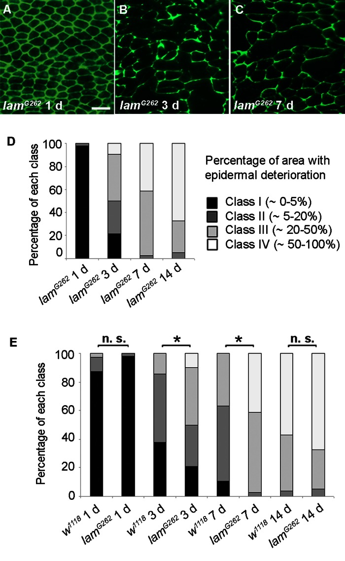
Figure 4.Epidermal Aging is Accelerated in Short-lived lamin Mutants(A-C) Anti-Fasciclin III immunofluorescence (green) of lamG262 epidermal whole mounts (n ≥ 10). Bar, 20 μm. A, 1 d. B, 3 d. C, 7 d. (D) Quantitative comparison of epidermal deterioration in lamG262 mutants and w1118 controls. All comparisons between different time points were siginificantly different using the Chi square test (p < 0.05). E, Quantitative comparison of epidermal deterioration in lamG262 mutants and w1118 controls. Significance indicators as in Figure 3D.