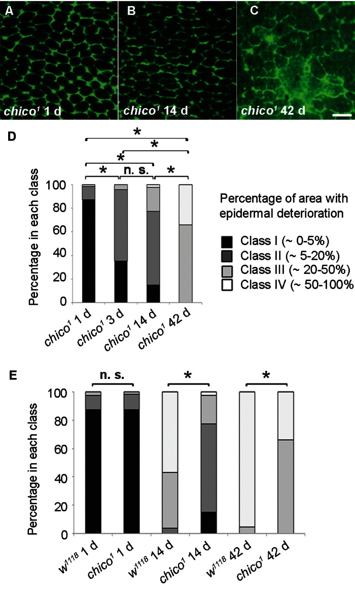
Figure 3.Decelerated Epidermal Aging in Long-lived chico Mutants(A-C) Anti-Fasciclin III immunofluorescence (green) of chico1 epidermal whole mounts (n ≥ 10). Bar, 20 μm. A, 1 d. B, 14 d. C, 42 d. (D) Quantification of chico1 epidermal deterioration with age. Asterisks, significant comparisons by Chi square test (p < 0.05); n. s., not significant. (E) Quantitative comparison of epidermal deterioration in chico1 mutants and w1118 controls. Significance indicators as in Figure 3D.