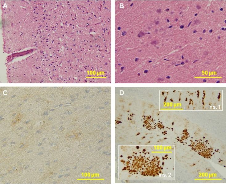
Figure 8.Histological and immunohistochemical examinations of brain tissues. (A) The cortex reveals no typical spongiform degeneration. (B) Several multicore plaques are present in the molecular layer of the cerebellum with a focal distribution. No pathology was seen in the pons and medulla. C: PrP immunostaining demonstrates weak staining with a synaptic pattern and occasional loose fine granular aggregates in the cerebral cortex. D: The molecular layer of the cerebellum shows a remarkable combination of strip-like staining (Ins. 1) and multicore plaques (Ins. 2). No immunostaining was seen in the pons and medulla.