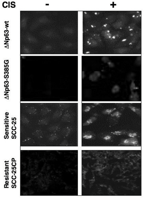
Figure 6.Immunofluorescence staining of LC3B expression in squamous carcinoma cells upon cisplatin exposureSets of wild type ΔNp63α/ΔNp63α-S385G cells and SCC-25/SCC-25CP cells were exposed to control media and 10μg/ml cisplatin for 24h. Cells were stained with a polyclonal antibody against MAP LC3α/β (1:100), and then photographed under fluorescent microscope.