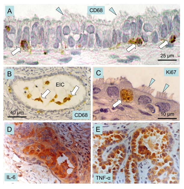
Figure 9.Macrophages, cytokines and ovarian cancer.
Epithelial inclusion cysts (EIC), showing infiltration of
the cyst wall (A) and lumen (B) by CD68 positive MDC
(arrows), ciliated cells (arrowheads), and Ki67 positive (arrow in C)
proliferating cells. Immunohistochemical staining of cancerous ovarian
tissues for IL-6. (D) and TNF-alpha (E) (x400). A-C adapted
from Ref. [111], © Elsevier, and D and E from
Ref. [113], © John Libbey Eurotext Ltd.