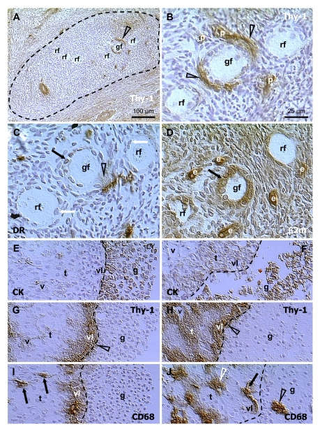
Figure 7.
Selection of secondary (A-D)
and preovulatory (dominant) follicles (E-F) in the adult human
ovary. Staining for Thy-1, HLA-DR (DR), MHC class I light chain (β2m),
cytokeratin 18 (CK) and CD68 of mature MDC, as indicated in panels. Dashed
line in (A) indicates an area exhibiting diminution of Thy-1
expression by stromal cells. (B), detail from (A). (C)
and (D) are semi-parallel sections to (B). Dashed line in (E-J),
follicular basement membrane. rf, resting follicles; gf, growing follicle;
p, pericytes; e, endothelial cells; v, microvasculature in theca interna
(t); vl, vascular layer adjacent to the follicular basement membrane; g,
granulosa layer. Details in text. Adapted from Ref. [70],
© Wiley-Blackwell.