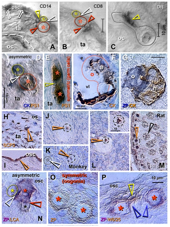
Figure 6.
Origin of new oocytes
(neo-oogenesis), primordial follicles, and SCP3 expression in adult human
and monkey ovaries (A-M), and oogenesis in adult rat ovaries (N-P).
(A) During asymmetric division (white arrowhead), the CD14 MDC
interact with both the OSC daughter (yellow arrowhead) and germ cell
daughter (red arrowhead). (B) T lymphocytes, however, interact with
the germ cell daughter only (red arrowheads). (C) Ameboid germ cells
(dotted line) migrating through the dense ovarian cortex (oc) are
accompanied by activated MDC (arrowhead). (D) Asymmetrically
dividing OSC produce a new PS1+ germ cell (red asterisk) and CK+ progenitor
cell (yellow asterisk). (E) In the tunica albuginea (ta) germ cells
(asterisks) symmetrically divide (arrowhead). (F) Capture of oocyte
(o) from the blood circulation by an arm (a) of granulosa cell nest (n)
lining the venule lumen (vl); e, endothelial cells. (G) Oocyte nest
assembly. (H) Segments of tunica albuginea (ta) in ovaries with
follicular renewal (early luteal phase) showed strong SCP3 expression of
mesenchymal (arrowheads) OSC precursors under ovarian surface (os). (I)
Staining of OSC (osc and arrowhead) was apparent in other segments - note
lack of staining of tunica albuginea under developed OSC. (J)
Postovulatory human ovaries showed staining of oocyte nucleoli (arrowhead)
in some primordial follicles. (K) In monkey ovaries, similar
staining of oocyte nucleoli in some primordial follicles was observed (red
vs. white arrowhead). (L) Staining of paired chromosomes oocyte was
observed in human ovaries (inset shows higher magnification). (M)
Adult rat testis (positive control) showed staining of condensed chromosomes
in spermatogonia (red arrowhead) and progression of meiotic division in
primary spermatocytes (black arrowhead). Oogenesis in adult rat ovaries is
initiated by asymmetric division of OSC (white arrowhead, N) showing
unstained OSC daughter (yellow asterisk) and ZP+ (magenta color) germ cell
daughter (red asterisk) accompanied like in human ovaries by a lymphocyte
(black asterisk and brown color). Symmetric division of ZP+ oogonia
(asterisks, O) follows, and is accompanied (P) by MDC (yellow
arrowhead). Blue arrowheads in (P) indicate association of primitive
granulosa cells with this process. ZP, zona pellucida; LCA, leukocyte
common antigen; W6/25, marker of rat MDC. Details in text. Adapted A-C from Ref. [57], © Blackwell Munksgaard, D-G from Ref. [35], © Antonin Bukovsky, H-M from Ref. [71], © Landes Bioscience, N-P from Ref. [72], © Landes Bioscience.