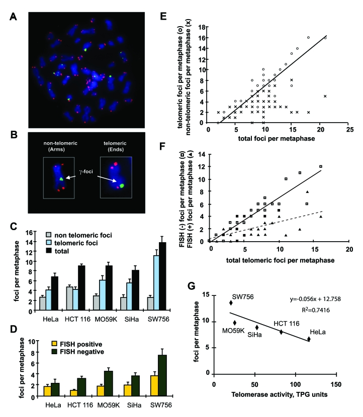
Figure 3.Distribution of γ-H2AX foci on metaphases of human tumor cells.(A) Metaphase spread of HCT116 cells stained for γ-H2AX (green) and telomeric DNA (red). (B)
Scoring of γ-H2AX foci as along chromatid
arms (Arms) or on chromatid ends (Ends). (C) The numbers of γ-H2AX foci in metaphases from five tumor cell
lines as noted. Foci are noted as non-telomeric (Arms, gray), telomeric
(Ends, blue), and total (black). (D) Telomeric γ-H2AX foci with (yellow) and without (green)
telomere FISH signal in the five tumor lines. At least 10 metaphases were
screened per data point in independent experiments. Error bars signify
standard errors. (E) Numbers of telomeric (open circles) and
non-telomeric (cross hatches) foci vs. the total numbers of γ-H2AX foci on the metaphase spreads of the five
tumor cell lines. The data from all five tumor cell lines was pooled for
this analysis. (F) Numbers of FISH negative (open squares) and FISH
positive telomeric (filled triangle) γ-H2AX
foci vs. total telomeric foci in all checked metaphases of the five tumor
cell lines. (G) Reverse correlation of the numbers of γ-H2AX foci and telomerase activity in the five
tumor cell lines. TPG is a total product generated corresponding to 600
molecules of telomerase substrate primers extended with at least four
telomere repeats [28].