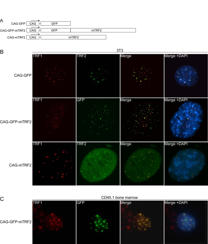
Figure 1.Lentiviral expression of mTRF2 and GFP-mTRF2.(A) Schematic of transgene constructs. GFP, mouse TRF2
(mTRF2) and a GFP-mTRF2 fusion were cloned into a lentiviral vector system [35] under the control of a CAG
promoter [38]. (B) Indirect
immunofluorescence of 3T3 cells. 3T3 control cells (top panel), 3T3 cells
transfected with GFP-mTRF2 and cells transfected with the mTRF2 construct
were stained with antibodies against mTRF1 or mTRF2. GFP was visualized by
autofluorescence. DNA has been stained with DAPI, and the merge of the red,
green and blue channels has been provided on the right. (C) Indirect
immunofluorescence of CD45.1 donor bone marrow cells. Cells were infected
with GFP-mTRF2 expressing lentiviruses and GFP-mTRF2 was visualized by GFP
autofluorescence. TRF1 was detected by a mTRF1 specific antibody, the DNA
was counterstained with DAPI And the merge of the three colors is
indicated.